Introduction and Context
Pyoderma gangrenosum (PG) is an uncommon neutrophilic dermatosis that typically presents with painful, rapidly progressive, sterile ulcerations. Clinical heterogeneity and frequent associations with systemic conditions—including inflammatory bowel disease (IBD), hematologic disorders, medications, and rare autoinflammatory syndromes—have long complicated diagnosis, reporting, and management. Despite the clinical burden and frequent need for systemic immunomodulation, no broadly accepted framework existed to categorize PG presentations in a way that informs clinical decision-making and research design.
In response to that gap, an international panel of experts used a modified Delphi process to create a standardized, descriptive classification framework for PG phenotypes and disease modifiers. The results were published as “Pyoderma Gangrenosum Phenotype Classification” (Gaurav et al., JAMA Dermatology, 2026). This article summarizes the consensus, explains why it matters, and highlights practical implications for clinicians and investigators.
Why this consensus was issued
– Clinical heterogeneity: PG presentations vary widely—ulcer morphology, anatomical sites, tempo, and associated systemic features differ across patients, affecting prognosis and response to therapy.
– Variable reporting and research heterogeneity: Studies and case series have used inconsistent definitions and classification systems, limiting meta-analyses and the generalizability of trial results.
– Treatment tailoring: Emerging biologic and targeted therapies (including anti–TNF agents, IL-1 and IL-17/IL-23 pathway inhibitors, and JAK inhibitors) require better phenotype-driven selection to optimize outcomes and safety.
– Need for a common language: Standardized phenotype categories can improve clinical guideline development, registry data quality, and stratified trial design.
The Delphi panel aimed to provide a descriptive, practical framework—not prescriptive treatment algorithms—so clinicians and researchers can classify PG presentations consistently.
New Guideline Highlights
Major outcomes of the consensus (Gaurav et al., 2026):
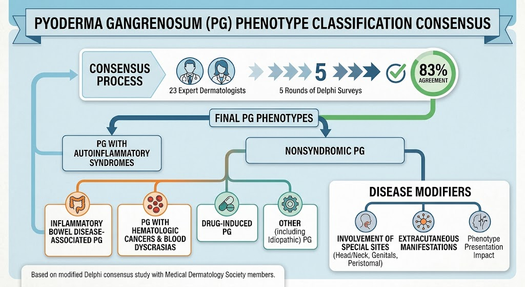
– Two overarching groups: (1) PG associated with autoinflammatory syndromes, and (2) nonsyndromic PG.
– Nonsyndromic PG subdivided into four phenotypes: (a) IBD-associated PG, (b) PG associated with hematologic cancers and blood dyscrasias, (c) drug-induced PG, and (d) other PG (including idiopathic cases).
– Disease modifiers recommended for routine documentation: involvement of special sites (head/neck, genitalia, or peristomal skin) and the presence of extracutaneous manifestations (e.g., sterile osteomyelitis, pulmonary involvement, systemic inflammatory features).
– Process and level of agreement: A 5-round modified Delphi survey of 23 board-certified dermatologists with PG expertise achieved 83% agreement on the final proposed framework; consensus threshold was predefined at ≥70%.
Key takeaways for clinicians:
– Adopt the descriptive phenotype labels when documenting PG cases and reporting in research.
– Use the phenotype and modifier framework as a guide to targeted diagnostic evaluation and multidisciplinary referrals (gastroenterology, hematology/oncology, rheumatology, geneticists).
– Consider phenotype when selecting therapies and designing trial eligibility criteria.
Updated Recommendations and Key Changes from Prior Approaches
Although previous efforts focused on diagnostic criteria (for example, the 2018 international Delphi defining diagnostic items for ulcerative PG; Maverakis et al., JAMA Dermatol. 2018), this 2026 consensus centers on phenotypic classification rather than diagnosis per se.
What’s new or different:
– Shift from morphology-only classification to etiology-anchored phenotypes: rather than only describing clinical appearance (ulcerative, pustular, bullous), the new framework explicitly ties phenotype to associated systemic conditions and putative triggers.
– Emphasis on modifiers: formal recognition that special anatomic sites and extracutaneous involvement materially modify disease course and therapeutic considerations.
– Standardized nomenclature for nonsyndromic subtypes, facilitating cohort stratification in registries and trials.
Why evidence supports these changes:
– Observational and case-series data have repeatedly shown differential associations and outcomes across PG linked to IBD, hematologic disease, and drugs—hence grouping by likely pathogenesis or driver supports precision in both diagnostic workup and therapy selection.
Topic-by-Topic Recommendations
Below are the major elements of the classification and recommended clinical actions tied to each phenotype. Note: the framework is descriptive; management decisions should continue to be individualized based on disease severity and patient comorbidities.
Phenotype structure (consensus categories):
– PG with autoinflammatory syndromes
– Nonsyndromic PG:
– Inflammatory bowel disease (IBD)–associated PG
– Hematologic cancers and blood dyscrasias–associated PG
– Drug-induced PG
– Other PG (including idiopathic)
Disease modifiers to document:
– Special site involvement: head/neck, genitalia/perineum, or peristomal skin
– Extracutaneous manifestations: sterile osteomyelitis, pulmonary sterile inflammation, systemic inflammatory signs, or others
Recommended diagnostic evaluation by phenotype (consensus-based, pragmatic guidance):
– All patients with suspected PG:
– Careful medication review for potential triggers
– Baseline laboratory panel: CBC with differential, CMP, inflammatory markers (ESR/CRP)
– Wound culture to rule out infection and guide antimicrobial stewardship
– Histopathology (punch or excisional biopsy) to exclude mimics—note that histologic features of PG are not pathognomonic
– If IBD-associated phenotype suspected:
– Screen for GI symptoms (diarrhea, hematochezia, abdominal pain), weight loss
– Consider fecal calprotectin and gastroenterology referral for colonoscopic evaluation
– Coordinate therapy to address both intestinal and cutaneous inflammation (communication with GI is essential)
– If hematologic-associated phenotype suspected:
– Review CBC for cytopenias or leukocytosis; peripheral smear
– Consider serum protein electrophoresis, immunophenotyping, and hematology referral if abnormal
– Recognize that treatment of the underlying hematologic disease can be critical to controlling PG
– If drug-induced phenotype suspected:
– Evaluate temporal relationship of new or increased-dose medications (notably granulocyte colony-stimulating factors, certain targeted therapies, and others reported in literature)
– Consider stopping the suspected drug if clinically feasible and monitor for improvement
– If autoinflammatory syndrome phenotype suspected:
– Screen for recurrent sterile neutrophilic inflammation, recurrent fevers, family history suggesting monogenic autoinflammatory disease
– Consider rheumatology/genetics referral and targeted genetic testing when indicated
– Special-site PG (head/neck, genital/perineal, or peristomal):
– Recognize increased morbidity; early multidisciplinary involvement (wound care, surgery, stoma care teams)
– Avoid unnecessary surgery or debridement without immunosuppression because of pathergy risk
Documentation and reporting recommendations:
– Record the assigned phenotype and any modifiers in the medical record and in registry reporting fields
– Report the criteria and evidence used to assign phenotype (e.g., active IBD confirmed by colonoscopy vs. historical diagnosis)
Expert Commentary and Insights
Committee views and contextual interpretation (paraphrased from the consensus process):
– A pragmatic, common-sense approach: Experts emphasized that the framework is intended as a practical taxonomy to harmonize reporting and research rather than as a rigid diagnostic algorithm.
– Balance between simplicity and clinical nuance: The panel aimed for categories that are broad enough to be widely applicable but specific enough to be clinically meaningful.
– Importance of modifiers: There was strong agreement that special anatomic sites and extracutaneous involvement change prognosis and management and should be recorded explicitly.
– Controversies and unresolved issues: Some panelists noted overlap between categories (for example, a patient with both IBD and a hematologic abnormality), raising the need for rules on how to prioritize or code overlapping etiologies in registries. The panel recommended documenting all relevant associations and allowing multi-label classification where needed.
– Future guidance: Experts urged that the classification be tested prospectively in registries and clinical trials to validate prognostic value and to refine phenotype-treatment links.
Practical Implications for Clinical Practice and Research
For clinicians:
– Use the phenotype labels to structure the diagnostic workup (targeted testing rather than exhaustive screening in every patient).
– Communicate phenotype and modifiers in consult notes and multidisciplinary discussions to guide therapy decisions.
– In special-site or extracutaneous disease, involve appropriate specialists early.
For researchers and guideline developers:
– Adopt the classification in registry data capture and clinical trial eligibility to allow stratified analyses (e.g., IBD-associated PG vs hematologic-associated PG).
– Test whether phenotype predicts response to specific therapies—this is a high-priority research need.
– Harmonize outcome measures and timing across phenotype-defined groups.
Potential impacts on therapy selection (evidence-informed suggestions):
– IBD-associated PG: consider therapies proven in IBD (e.g., anti–TNF agents) when appropriate and coordinate combined GI–dermatology care.
– Hematologic-associated PG: treat underlying hematologic disease promptly and involve hematology; targeted oncology/hematology therapies may be required.
– Drug-induced PG: stopping the offending agent may be sufficient; immunosuppression reserved for persistent lesions.
– Autoinflammatory syndromes: targeted agents (IL-1 inhibitors, etc.) may be considered in select cases under specialist guidance.
Note: These management considerations echo existing literature and practice patterns, but the consensus classification itself does not replace individualized therapeutic decision-making.
Next Steps and Research Directions
– Prospective validation: Implement the framework in prospective registries (dermatology and multidisciplinary) to test associations with outcomes and treatment response.
– Trial stratification: Use phenotype labels in trial design to ensure balanced or stratified randomization and to enable phenotype-specific efficacy analyses.
– Refinement of categories: As new evidence accumulates—especially around molecular and genetic drivers—phenotype definitions may be refined or expanded.
– Standardized data elements: Encourage societies and registries to adopt recommended data fields (phenotype, modifiers, supporting evidence) to harmonize reporting.
Patient Vignette
Anna, a 44-year-old woman with a history of ulcerative colitis, presents with a rapidly enlarging, painful ulcer on her shin. Workup finds active intestinal inflammation on colonoscopy. Her case would be classified as “nonsyndromic PG — IBD-associated phenotype,” with no special-site modifier. Practical implications: involve GI and dermatology, consider systemic therapy that can address both cutaneous and intestinal inflammation, and document phenotype for future registry reporting.
References
– Gaurav A, Gregoire S, Xia E, McIntosh BA, Alavi A, Bolognia J, Cowen E, Dominguez AR, Fernandez AP, Fivenson D, Heffernan M, Huang W, Kaffenberger B, Madigan L, Mauskar M, Means AD, Nelson C, Patsatsi A, Pugliese D, Rojek NW, Rosenbach M, Shaigany S, Shinkai K, Smith G, Swerlick R, Ortega-Loayza A, Mostaghimi A. Pyoderma Gangrenosum Phenotype Classification. JAMA Dermatol. 2026 Aug 5. PMID: 42555014. https://pubmed.ncbi.nlm.nih.gov/42555014/
– Maverakis E, Ma C, Shinkai K, et al. Diagnostic criteria of ulcerative pyoderma gangrenosum: a Delphi consensus of international experts. JAMA Dermatol. 2018;154(5):561–566. doi:10.1001/jamadermatol.2017.6321
(Additional background references on PG epidemiology and management can be consulted in leading reviews and specialty resources such as UpToDate and major dermatology textbooks.)
Conclusion
The 2026 expert consensus provides the first widely endorsed, descriptive phenotype classification for pyoderma gangrenosum. By defining phenotype groups and disease modifiers, the framework offers a practical taxonomy that can improve clinical evaluation, harmonize reporting, and enable phenotype-driven research and trials. Adoption by clinicians, registries, and trialists will be essential for validating and refining the system and ultimately for improving outcomes in this challenging disease.