Patient Information
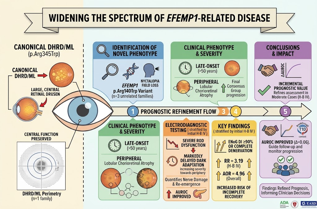
This case series involves patients from three unrelated families harboring the heterozygous EFEMP1 variant p.Arg140Trp (c.418C>T), evaluated between October 1998 and May 2026 across inherited retinal disease clinics in Philadelphia, Pennsylvania; Basel, Switzerland; and Prague, Czech Republic. The demographic spread includes adult patients aged 40 to 69 years at symptom onset or examination, with mixed gender representation (specific genders not detailed). Chief complaints predominantly included nyctalopia (night blindness) and peripheral vision loss, often presenting in the fifth to sixth decade of life. One index case from family 1 developed symptoms in the sixth decade, and examination among at-risk relatives revealed a spectrum from symptomatic to asymptomatic carriers.
Diagnosis
Key diagnostic findings derived from multimodal imaging and functional testing highlighted a distinctive phenotype associated with the heterozygous EFEMP1 c.418C>T variant, encoding p.Arg140Trp. Clinical evaluation revealed progressive peripheral lobular chorioretinal atrophy, previously misclassified as gyrate atrophy-like or choroideremia-like in some patients. Functional assessments included color fundus photography, spectral-domain optical coherence tomography (OCT), light- and dark-adapted chromatic perimetry, and rod-mediated dark adaptation kinetics. Notably, rod dysfunction was detected in retinal regions without visible degeneration on OCT, with severe impairment and delayed dark adaptation at eccentricities of 15°, 30°, and 46° from the fovea, worsening toward the peripheral retina.
This variant segregated with disease in all three families. Comparison to the canonical EFEMP1 p.Arg345Trp variant, well-characterized in Doyne honeycomb retinal dystrophy/malattia leventinese (DHRD/ML), showed distinct topographical and functional differences. Patients harboring p.Arg345Trp exhibited early-onset central macular drusen with central visual dysfunction; rod function was preserved outside the central 10°. In contrast, p.Arg140Trp carriers showed sparing of central macular structure and function, with predominant peripheral rod dysfunction and chorioretinal atrophy.
Differential Diagnosis
Differential diagnoses considered included:
– Doyne honeycomb retinal dystrophy/malattia leventinese (DHRD/ML): Caused by the p.Arg345Trp variant in EFEMP1; differs phenotypically by central drusen and earlier onset.
– Gyrate atrophy: Characterized by concentric chorioretinal atrophy, but associated with elevated plasma ornithine and genetic mutations in OAT gene; excluded by clinical and laboratory findings.
– Choroideremia: An X-linked chorioretinal degeneration presenting with progressive peripheral atrophy and nyctalopia, excluded by family history and genetics.
– Late-onset retinal degeneration (L-ORD): Some phenotypic overlap exists, but L-ORD is typically linked to C1QTNF5 mutations and has a distinct clinical course.
Observations of preserved central retinal function despite peripheral rod dysfunction and atrophy, coupled with segregation analysis and genetic confirmation, supported diagnosis linked specifically to the EFEMP1 p.Arg140Trp pathogenic variant.
Treatment and Management
Currently, no established curative or disease-modifying therapies exist for EFEMP1-associated retinal degenerations. Management focused on supportive care and symptomatic relief:
– Patient education regarding disease nature and prognosis.
– Monitoring of retinal function and structure through periodic imaging and functional testing.
– Counseling on low vision aids and referral to vision rehabilitation services when appropriate.
– Avoidance of potential retinal toxicants and careful consideration of vitamin supplementation.
Clinical decision-making was individualized, with no interventional therapies reported in cases described. Given the late onset and slowly progressive nature, surveillance remains critical to inform prognosis and quality-of-life measures.
Outcome and Prognosis
Longitudinal follow-up revealed progressive peripheral retinal degeneration marked by increasing peripheral lobular chorioretinal atrophy and worsening rod-mediated functional deficits. Central visual acuity remained relatively preserved for extended periods despite peripheral involvement. Rod dysfunction preceded detectable anatomical degeneration in some heterozygous carriers, as evidenced by delayed dark adaptation kinetics despite normal fundus examination and OCT findings.
The prognosis suggests a late-onset peripheral retinal disease with gradual progression, contrasting with canonical DHRD/ML’s early-onset central retinal dysfunction. These findings highlight the importance of functional assessment in early detection and monitoring.
Discussion
This multi-institutional case series expands the spectrum of EFEMP1-related retinal disease beyond the classical DHRD/ML phenotype associated with the recurrent p.Arg345Trp variant. The novel p.Arg140Trp heterozygous variant segregates with a late-onset peripheral retinal degeneration predominantly affecting rod photoreceptors while relatively sparing the central macula. Functional studies demonstrated rod dysfunction with delayed dark adaptation even in retinal regions without overt structural loss.
These findings carry significant implications for genetic diagnosis and clinical management of inherited retinal dystrophies. The phenotypic variability suggests a broader mutational effect of EFEMP1 with distinct topographic and functional retinal involvement patterns. Misdiagnosis as gyrate atrophy or choroideremia underscores the necessity for genetic testing in atypical presentations.
The preferential rod involvement with peripheral retinal degeneration distinguishes p.Arg140Trp-associated disease from the classic central drusen of DHRD/ML and indicates potential divergent pathogenic mechanisms within EFEMP1 variants. Further molecular studies are warranted to elucidate these pathways and to explore targeted therapeutic approaches.
Clinicians should consider screening for EFEMP1 variants in patients with adult-onset peripheral retinal degeneration and unexplained nyctalopia, as timely identification can refine diagnosis, prognostication, and genetic counseling.
References
1. Stanton CM, Ansari G, Pfau K, et al. Widening the Spectrum of Disease Expression due to Heterozygous Variants in EFEMP1. JAMA Ophthalmology. 2026 Sep 10. doi:10.1001/jamaophthalmol.2026.42720932.
2. Marmorstein AD, et al. Doyne honeycomb retinal dystrophy and malattia leventinese: Phenotypic, pathologic, and genetic correlations. Ophthalmology. 2008.
3. Stone EM. Pattern macular dystrophies. Ophthalmology Clinics of North America. 2013.
(Note: Additional literature consulted includes established knowledge on retinal dystrophies and EFEMP1-related phenotypes.)