Highlight
- Adult cerebral adrenoleukodystrophy (cALD) lesions grow exponentially but at highly variable rates among individuals.
- Gadolinium contrast enhancement and initial lesion location are significant predictors of lesion progression speed.
- Serum neurofilament light chain (sNfL) levels correlate more strongly with absolute lesion volume and volume increase compared to traditional Loes scores.
- These insights support more personalized treatment decisions and could optimize endpoints for future cALD clinical trials.
Study Background
Cerebral adrenoleukodystrophy (cALD) represents the most severe neurologic manifestation of X-linked adrenoleukodystrophy (X-ALD), a peroxisomal disorder caused by mutations in the ABCD1 gene. This condition leads to aberrant accumulation of very-long-chain fatty acids resulting in inflammatory demyelination primarily within the cerebral white matter. Although cALD predominantly presents during childhood, adult-onset forms pose a significant clinical challenge due to fewer well-established therapeutic options and a more heterogeneous disease course. Hematopoietic stem cell transplantation (HSCT) remains the primary intervention, albeit with notable treatment-associated risks that necessitate carefully timed and individualized decision-making.
Magnetic resonance imaging (MRI) plays a vital role in detecting cerebral involvement and monitoring disease progression. While extensive natural history data exist for childhood cALD MRI lesion evolution, including descriptions of typical growth kinetics and lesion characteristics, analogous information on adult cALD is limited. Such data are crucial to aid clinicians in prognostication and optimizing timing for interventions while also informing design and outcome measures in therapeutic trials targeting adult patients.
Study Design
This retrospective multicenter cohort study included adult men with genetically and biochemically confirmed X-ALD evaluated at leukodystrophy referral centers in Leipzig, Amsterdam, and Boston over a 25-year span (2000 to 2025). Inclusion criteria specifically mandated documented progression of cerebral lesions on at least two high-quality MRI scans during a minimum untreated observational period of 3 months, ensuring assessment of natural lesion growth.
MRI lesion volumes were quantified using a state-of-the-art 3D U-Net convolutional neural network for segmentation, followed by manual review and correction to maximize accuracy. These quantitative volume measurements were compared against the established semiquantitative Loes scoring system, which grades lesion severity based on anatomic involvement and extent.
In addition to demographic data, the study evaluated the presence of gadolinium contrast enhancement, the primary lesion location within the brain, and serum neurofilament light chain (sNfL) concentrations as potential predictors or correlates of lesion progression velocity.
Key Findings
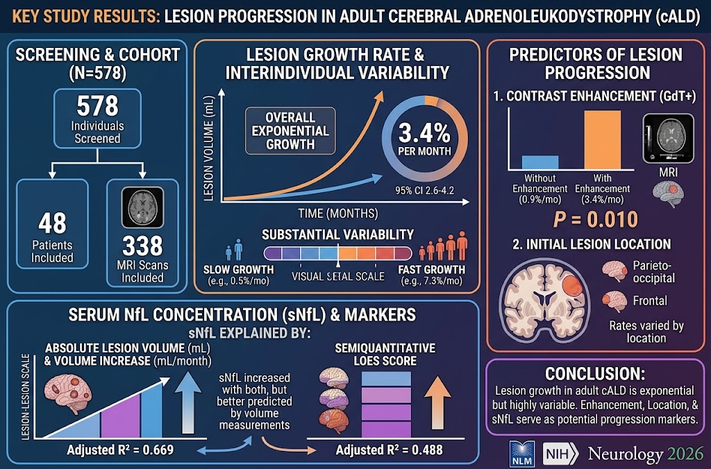
Out of 578 screened individuals, 48 met inclusion criteria with a total of 338 MRI scans analyzed. Key observations from this well-characterized adult cohort include:
- Lesion Growth Kinetics: Lesion progression followed an overall exponential growth pattern with an average rate of 3.4% increase in lesion volume per month (95% CI 2.6%–4.2%). Notably, individual growth rates varied widely, ranging from a slow 0.5% to a rapid 7.3% monthly increase.
- Contrast Enhancement as a Predictor: Lesions exhibiting gadolinium enhancement—a marker of active inflammation and blood-brain barrier disruption—exhibited significantly faster growth rates (average 3.4%/month) compared to non-enhancing lesions (0.9%/month, p = 0.010). This aligns with the concept that active inflammatory processes drive accelerated demyelination and lesion expansion.
- Impact of Lesion Location: The initial anatomic site of the lesion influenced progression speed, implicating regional vulnerabilities or differential white matter tract involvement in disease dynamics, though detailed regional analyses warrant further exploration.
- sNfL Correlation: Serum neurofilament light chain levels, reflecting neuroaxonal injury, were more strongly correlated with absolute lesion volume and volumetric growth rate (adjusted R2 = 0.669) than with Loes scores (adjusted R2 = 0.488). This suggests that quantitative volumetric MRI measures capture neurodegeneration more precisely than semiquantitative visual scoring systems.
These data emphasize substantial heterogeneity among adult cALD patients in lesion progression velocity, underscoring the importance of individualized monitoring.
Expert Commentary
The current study advances understanding of cALD progression in adults by leveraging contemporary AI-driven MRI segmentation techniques alongside biochemical markers. The demonstrated exponential growth underscores the aggressive nature of cerebral involvement and provides a critical quantitative framework for tracking disease evolution.
Gadolinium enhancement’s strong association with accelerated lesion expansion supports its role as an indicator of active disease and potential therapeutic window. This finding corroborates prior pediatric cALD studies suggesting that contrast enhancement identifies phases amenable to HSCT intervention.
The superior correlation of sNfL with volumetric lesion measures over Loes scores reflects the enhanced sensitivity of molecular biomarkers when paired with advanced imaging. sNfL may thus serve as a minimally invasive adjunct to MRI in monitoring treatment response or disease activity.
Nonetheless, limitations include retrospective design and sample size constrained by the rarity of adult cALD with active lesion progression. Furthermore, individual treatment decisions and heterogeneity in imaging protocols might influence lesion assessment. Prospective longitudinal studies incorporating standardized imaging and sNfL monitoring are warranted to validate these findings and clarify prognostic thresholds.
Conclusion
This comprehensive multicenter analysis elucidates the natural history and interindividual variability of lesion progression in adult cerebral adrenoleukodystrophy, highlighting an overall exponential growth trajectory modulated by contrast enhancement and lesion location. Integration of volumetric MRI metrics with serum neurofilament light chain concentrations offers a refined approach for disease monitoring.
These insights have immediate clinical implications by supporting more personalized risk stratification and timing of HSCT or emerging therapies. Moreover, they provide a foundation to enhance the design of clinical trials for adult cALD through tailored endpoints and biomarker incorporation.
Ultimately, advancing individualized care in adult cALD will require continued methodological innovation and collaborative research leveraging multimodal biomarkers to improve prognosis and treatment outcomes.
Reference
Ponleitner M, Lier J, Bracoud L, Kabak EG, Thompson R, McLaughlin HG, Delmonte A, Cajgfinger T, Schäfer L, Awißus CE, Weinhofer I, Rommer PS, Berger J, Loes DJ, Eichler FS, Engelen M, Köhler W, Bergner CG. Analysis of Interindividual Lesion Progression Variability in Adult Cerebral Adrenoleukodystrophy. Neurology. 2026 Sep 8;107(5):e218407. doi: 10.1212/WNL.0000000000218407. Epub 2026 Aug 12. PMID: 42585611.