Highlight
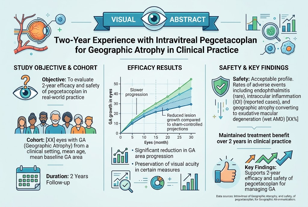
This two-year real-world observational study demonstrates that intravitreal pegcetacoplan therapy reduces the growth rate of geographic atrophy (GA) by approximately 41%, with sustained efficacy in the second year. Treatment was well-tolerated in 77 eyes from 58 patients, with a manageable injection frequency and consistent monitoring using swept-source optical coherence tomography angiography (SS-OCTA). Visual acuity showed a modest decline, reflecting the natural course of late-stage AMD despite slowed lesion progression.
Study Background
Geographic atrophy (GA) is the advanced, non-exudative form of age-related macular degeneration (AMD) characterized by progressive retinal pigment epithelium and photoreceptor loss, leading to irreversible vision impairment. GA is a significant cause of central visual loss in elderly populations, with no established therapies until recently. The complement cascade, particularly C3 activation, has been implicated in GA pathogenesis, prompting the development of complement inhibitors such as pegcetacoplan, an intravitreal C3 inhibitor. Controlled clinical trials showed promise in slowing GA lesion growth, but long-term real-world data assessing efficacy, dosing, and safety remain sparse.
Study Design
This investigation was a retrospective interventional case series involving 58 patients (77 eyes) with symptomatic GA secondary to AMD treated at a single center under clinical practice conditions between April 2023 and December 2025. Inclusion criteria required confirmed GA with measurable lesions. Patients received intravitreal injections of 15 mg pegcetacoplan, administered at an average interval of 1.3 months, totaling about 18 injections per eye over two years. All subjects participated in a prospective imaging arm utilizing swept-source optical coherence tomography angiography (SS-OCTA) before and during treatment to evaluate lesion morphology, GA size, and vascular status, including detection of non-exudative macular neovascularization (MNV) and monitoring for exudation onset. Key endpoints included GA growth rate calculated by square-root lesion size change, best-corrected visual acuity (BCVA) changes, and safety observations.
Key Findings
Among 72 eyes with complete 2-year follow-up and measurable GA, the mean square-root GA growth rate decreased from a baseline of 0.33±0.21 mm/year to 0.19±0.11 mm/year after treatment, representing a significant 41% reduction (p<0.001). Notably, 71 of these eyes showed either a stable or further decreased GA growth rate in the second year, indicating sustained or cumulative treatment efficacy. Only one eye exhibited increased growth rate and required adjunctive anti-VEGF therapy for concomitant exudative MNV.
There was no significant difference in GA growth inhibition based on lesion location, with comparable responses observed in eyes with foveal versus non-foveal GA (p≥0.99). The mean BCVA declined modestly from 63±14 Early Treatment Diabetic Retinopathy Study (ETDRS) letters at baseline to 55±16 letters at 2 years (p<0.001), consistent with natural disease progression despite treatment. BCVA decline did not significantly differ between foveal and non-foveal lesions (p=0.14).
A positive correlation was demonstrated between the baseline GA growth rate and the extent of growth inhibition over one and two years (r=0.33, p=0.02 and r=0.40, p=0.007, respectively), suggesting patients with faster initial progression may experience greater therapeutic benefit.
The treatment regimen was tolerable, with an average of 18 injections administered per eye over two years and no major safety concerns reported. SS-OCTA imaging enabled detailed surveillance of lesion evolution and early detection of neovascular complications.
Expert Commentary
These real-world data validate and extend findings from controlled clinical trials on pegcetacoplan’s efficacy in slowing GA progression. The observed 41% reduction in GA growth rate aligns well with prior phase 3 trial outcomes, demonstrating that complement C3 inhibition can effectively decelerate retinal degeneration attributable to AMD.
The moderate visual acuity decline despite lesion growth attenuation underscores the multifactorial nature of vision loss in GA, involving photoreceptor function and neural remodeling beyond lesion size alone. Continuous monitoring with advanced imaging such as SS-OCTA proves critical in optimizing individualized treatment intervals and detecting coexistent neovascularization requiring adjunct therapies.
Limitations include the retrospective design and absence of a randomized control group. Also, clinical practice injection intervals varied somewhat from trial protocols, which might influence outcome comparability. Additionally, longer-term data beyond two years will be essential to confirm sustained benefit and safety.
Conclusion
In clinical practice, intravitreal pegcetacoplan administered approximately every 1.3 months is associated with a significant and durable 41% reduction in GA growth rate over two years in patients with AMD. While visual acuity decline persists, the slowed anatomic progression offers a meaningful therapeutic advance for a previously untreatable condition. These findings support pegcetacoplan as a valuable treatment option for GA, with continued research required to refine treatment protocols and assess long-term functional outcomes.
Funding and Clinical Trials Registration
The original study did not specify funding sources or clinical trial registry numbers. Readers are advised to consult the primary publication for detailed disclosures.
References
1. Jaffe GJ, Westby K, Csaky KG, et al. C5 Inhibitor Avacincaptad Pegol for Geographic Atrophy due to Age-Related Macular Degeneration: A Randomized Pivotal Phase 3 Trial. Ophthalmology. 2021;128(4):576-586.
2. Liao DS, Grossi FV, El Mehdi D, et al. Complement C3 Inhibitor Pegcetacoplan for Geographic Atrophy Secondary to Age-Related Macular Degeneration: A Randomized Phase 2 Trial. Ophthalmology. 2020;127(2):186-195.
3. Holz FG, Sadda SR, Staurenghi G, et al. Imaging Protocols in Geographic Atrophy: A consensus. Ophthalmology Retina. 2020;4(4):343-349.
4. Shen M, Lam WZ, Berni A, et al. Two-Year Experience with Intravitreal Pegcetacoplan for the Treatment of Geographic Atrophy in Clinical Practice. American journal of ophthalmology. 2026 Sep 11. PMID: 42727684.