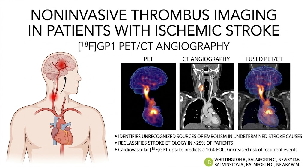
Highlight
– [18F]GP1 PET/CT imaging directly visualizes activated platelet thrombi in acute ischemic stroke patients.
– Identification of thrombus origin leads to reclassification of stroke subtype in over 25% of cases.
– Detection of cardiovascular thrombus correlates with a significant increase in recurrent cerebrovascular events.
– This diagnostic advance facilitates mechanism-based stroke classification and targeted prevention.
Study Background
Acute ischemic stroke remains a leading cause of morbidity and mortality worldwide. Accurate identification of the source of thromboembolism is crucial for selecting optimal secondary prevention strategies to reduce recurrence. Traditional stroke classification systems often rely on indirect, circumstantial evidence such as clinical features, vascular imaging, and cardiac evaluations. These approaches may leave nearly a third of strokes labeled as cryptogenic or of undetermined etiology, challenging effective management. Consequently, there is an unmet clinical need for technology that can noninvasively and directly identify thrombus formation to clarify stroke mechanisms.
Study Design
This prospective single-center cohort study enrolled 100 patients presenting with acute ischemic stroke within 21 days of symptom onset. In addition to comprehensive standard-of-care stroke investigations including clinical assessment, vascular imaging, and cardiac evaluation, patients underwent hybrid imaging using a novel radiotracer, fluorine-18 glycoprotein IIb/IIIa inhibitor ([18F]GP1), combined with positron emission tomography (PET) and computed tomography angiography (CTA). The [18F]GP1 tracer specifically binds to activated platelet glycoprotein IIb/IIIa receptors, enabling visualization of platelet-rich thrombi in vivo.
Stroke etiology was independently established by expert stroke physicians blinded to imaging results, employing the causative classification system. Separately, blinded readers analyzed the PET/CTA images to identify sites of thrombus uptake in cardiovascular structures without knowledge of clinical data or stroke subtype classification. The main objectives were to determine the ability of [18F]GP1 PET/CTA to detect thrombus source, impact stroke classification, and predict recurrence of stroke or transient ischemic attacks (TIA) during follow-up.
Key Findings
The study found that cardiovascular thrombus with [18F]GP1 uptake was detected in 63 of 100 patients (63%). This imaging evidence led to a change in stroke subtype classification in 26 patients (26%), notably improving diagnostic clarity.
Among the 41 patients initially classified with stroke of undetermined cause, the thrombus source was revealed by PET/CTA in 18 cases (44%). These thrombi included nonstenotic carotid atherothrombosis, native cardiac valve thrombosis, and paradoxical embolism, all of which were not previously identified through conventional workup. This underscores the value of direct thrombus imaging in revealing otherwise occult embolic sources.
During a median follow-up period of 613 days (interquartile range, 251-788 days), 14 patients experienced recurrent stroke or TIA. Remarkably, 13 of these 14 patients (93%) had evidence of cardiovascular thrombus detected by [18F]GP1 PET/CTA at baseline. Statistical analysis demonstrated that presence of PET/CTA thrombus uptake predicted a 10.4-fold increased hazard of recurrent cerebrovascular events (95% CI, 1.35-79.40; P=0.024), indicating strong prognostic value.
Expert Commentary
This study represents a major advance in the stroke diagnostic paradigm by shifting from inference-based assessments to direct visualization of thrombus formation, enhancing etiologic precision. The use of [18F]GP1 PET/CT allows detection of active platelet aggregation at embolic sources that traditional imaging might miss, such as nonstenotic plaques or small cardiac thrombi.
However, as a single-center observational study, these findings require validation in broader, multi-center cohorts with diverse populations. Limitations include the availability, cost, and logistics related to PET radiotracer production and imaging. Further research should evaluate the impact of this imaging approach on clinical decision-making, treatment adjustments, and long-term outcomes.
The biological plausibility is strong, given the selective binding of [18F]GP1 to activated platelet receptors, providing a mechanistic link between thrombus detection and embolic risk. These insights may inform future guidelines on stroke evaluations and personalized secondary prevention strategies.
Conclusion
Noninvasive thrombosis imaging with [18F]GP1 PET/CT is a promising innovation that enhances the detection of active thrombus in acute ischemic stroke patients, providing unprecedented etiologic clarity. By revealing the cardiovascular source of emboli in nearly half of undetermined strokes, this technology has the potential to transform stroke classification, guide targeted interventions, and predict recurrent cerebrovascular events. Integration of mechanism-based stroke diagnosis could improve patient outcomes and reduce the burden of stroke recurrence.
Further multi-center studies are needed to confirm these findings and to evaluate cost-effectiveness before routine clinical adoption.
Funding and Trial Registration
This study was registered at ClinicalTrials.gov with identifier NCT05636748. Details on funding sources were not specified in the abstract.
References
Whittington B, Balmforth C, Giaj Levra A, et al. Noninvasive Thrombus Imaging in Patients With Ischemic Stroke. Circulation. 2026 Sep 1; PMID: 42677492. Available from: https://pubmed.ncbi.nlm.nih.gov/42677492/
Additional relevant literature on stroke classification and imaging modalities can be referenced from current guidelines such as the American Heart Association/American Stroke Association for comprehensive context.