Highlight
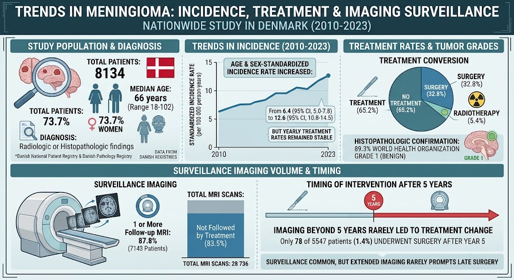
– The incidence of meningioma in Denmark nearly doubled from 2010 to 2023, rising from 6.4 to 12.6 per 100,000 person-years.
– Despite this increase, treatment rates via surgery or radiotherapy remained stable, with 65% of patients receiving no intervention.
– Follow-up magnetic resonance imaging (MRI) was extensively utilized, but over 80% of scans did not lead to treatment changes.
– Imaging conducted beyond five years post-diagnosis rarely prompted intervention, suggesting potential for reducing surveillance intensity through risk-adapted protocols.
Study Background
Meningiomas are the most common primary intracranial tumors, generally benign and often indolent in nature. Despite their typically slow-growing behavior, the worldwide reported incidence rates of meningioma have been rising. Advances in neuroimaging have improved tumor detection, including incidental findings, raising concerns regarding overtreatment and the necessity of extensive diagnostic surveillance. Given that many patients remain asymptomatic and untreated, the clinical burden of frequent neuroimaging follow-ups—both in terms of healthcare resources and patient anxiety—is substantial. A critical gap exists in understanding whether current follow-up strategies meaningfully guide treatment decisions or could be optimized for efficiency without compromising patient outcomes.
Study Design
This quality improvement study utilized nationwide Danish registries—the Danish National Patient Registry and Danish Pathology Registry—to identify adults diagnosed with meningioma, either radiologically or histopathologically confirmed, between January 1, 2010, and December 31, 2023. Follow-up extended through December 31, 2024. The study quantified temporal trends in meningioma incidence and subsequent treatment, defined as surgery or radiotherapy. Surveillance imaging volumes were analyzed, focusing on magnetic resonance imaging (MRI), detailing the timing and frequency of scans relative to treatment events.
Key Findings
Among 8,134 meningioma patients included, the median age was 66 years with a predominance of women (73.7%). The standardized incidence rate rose markedly from 6.4 (95% CI, 5.0-7.8) to 12.6 (95% CI, 10.8-14.5) per 100,000 person-years over the study period. However, the proportion of patients receiving active treatment remained constant yearly: 32.8% underwent surgery, 5.4% received radiotherapy, and the majority (65.2%) remained untreated.
Histopathology confirmed World Health Organization (WHO) grade 1 meningiomas in 89.3% of surgically treated cases, reflecting the mostly benign nature of the tumors diagnosed.
Regarding surveillance, 87.8% of patients had at least one follow-up MRI scan, culminating in 28,736 scans overall. Importantly, 83.5% of these scans were not followed by any change in treatment strategy. Surveillance imaging beyond five years after diagnosis represented a significant fraction of total scans but infrequently led to interventions; only 1.4% of patients underwent surgery after five years, suggesting a low likelihood of delayed progression requiring treatment.
Expert Commentary
This comprehensive nationwide dataset provides valuable insights into meningioma management in an era of increasing detection. The doubling of incidence likely reflects enhanced neuroimaging utilization and improved diagnostic sensitivity for incidental meningiomas rather than a true biologic surge. The stable treatment rates underscore that most tumors detected are clinically indolent, supporting conservative management approaches.
Extensive follow-up imaging appears to offer limited benefit in guiding treatment decisions, particularly for patients with confirmed low-grade tumors who remain asymptomatic. Prolonged imaging surveillance beyond five years rarely results in changes to patient management, highlighting an opportunity to safely de-escalate monitoring in selected low-risk patients.
Despite these strengths, the study’s reliance on registry data may limit granularity regarding tumor size, location, and symptomatology, factors critical for individualized risk stratification. Furthermore, patient-reported outcomes and quality-of-life data were not incorporated, which are essential in evaluating the clinical impact of surveillance strategies.
Conclusions
This quality improvement study reveals a significant increase in meningioma diagnoses in Denmark over 13 years without a corresponding rise in treatment interventions. The prevalent use of MRI surveillance often yields no changes to management, particularly beyond five years after diagnosis. These findings advocate for the development and implementation of risk-adapted follow-up protocols that minimize unnecessary imaging, reduce healthcare costs, and lessen patient burden while maintaining vigilant detection of clinically relevant progression.
Funding and Clinical Trials Registry
The original study did not report specific funding sources or clinical trial registration. Future research should aim to explore prospective, multicenter trials evaluating tailored surveillance algorithms based on tumor characteristics and patient risk profiles.
References
1. Korshøj AR, Engberg H, Haldrup M, et al. Trends in Meningioma Incidence, Treatment, and Imaging Surveillance. JAMA Oncol. 2026;doi:10.1001/jamaoncol.42623066.
2. Ostrom QT, Gittleman H, Xu J, et al. CBTRUS Statistical Report: Primary Brain and Other Central Nervous System Tumors Diagnosed in the United States in 2014-2018. Neuro Oncol. 2021;23(Suppl 2):iii1-iii105.
3. Goldbrunner R, Minniti G, Preusser M, et al. European Association of Neuro-Oncology (EANO) guideline on the diagnosis and management of meningiomas. Neuro Oncol. 2021;23(11):1821-1834.
4. Lee DS, Park SH, Cho JH, et al. Natural history of asymptomatic incidental meningiomas. J Neurosurg. 2022;136(4):1240-1247.
5. Sarmiento JM, Butler WE, Kornblum HI, et al. Imaging surveillance and surgical management of incidental meningiomas. Neurosurg Focus. 2020;49(6):E8.