Highlights
Normative modeling analysis of 631 participants reveals that conventional group-based approaches mask substantial individual-level heterogeneity in traumatic brain injury (TBI), with no more than 23% of patients sharing extreme deviations in any single brain region despite significant case-control findings. Median morphologic deviations increase progressively with injury severity, from 9 in mild cases to 22 in severe cases, demonstrating the value of individualized neuroanatomical assessment for advancing personalized TBI management.
Background: The Challenge of TBI Heterogeneity
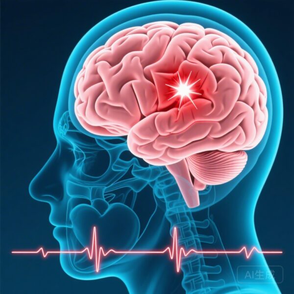
Traumatic brain injury represents one of the most complex neurological conditions in modern medicine, affecting approximately 69 million individuals worldwide annually. Despite decades of research, clinical management of TBI remains challenging, in large part because no two brain injuries are alike. The heterogeneity of TBI—encompassing diverse injury mechanisms, locations, severities, and individual biological responses—creates substantial obstacles for developing standardized diagnostic criteria, prognostic algorithms, and therapeutic interventions.
Traditional neuroimaging research has relied predominantly on case-control study designs, comparing groups of TBI patients against healthy controls to identify statistically significant differences. While such approaches have generated important insights into the neuroanatomical consequences of brain injury, they inherently compress individual variation into group-level summaries. This averaging effect can obscure the true complexity of TBI-related brain changes, potentially missing clinically relevant individual deviations that might inform personalized treatment strategies.
The study by Mitchell and colleagues addresses this fundamental limitation by introducing normative modeling as an alternative analytical framework for characterizing brain morphology in TBI. Rather than asking whether TBI patients differ from healthy individuals as a group, normative modeling asks how each individual deviates from expected patterns derived from healthy reference populations. This paradigm shift enables detection of participant-specific abnormalities that conventional group comparisons may overlook entirely.
Study Design and Methodology
The research team conducted a comprehensive analysis leveraging data from the Enhancing NeuroImaging Genetics through Meta-Analysis (ENIGMA) Consortium Adult Moderate-to-Severe Traumatic Brain Injury working group. The study included 631 magnetic resonance imaging (MRI) examinations, comprising 407 patients with documented TBI and 224 age- and sex-matched healthy controls, enabling robust comparative analysis across the full spectrum of injury severity.
The analytical approach employed two complementary methodologies. First, traditional case-control analyses utilized linear models examining cortical thickness and subcortical volumes, with statistical adjustments for age, sex, imaging site, and intracranial volume to ensure valid comparisons. Second, normative modeling was conducted through the Predictive Clinical Neuroscience Portal, employing Bayesian linear regression with likelihood warping to estimate individual deviations from expected neuroanatomical distributions.
Brain morphology was quantified using established neuroimaging protocols, with cortical thickness measurements derived from the Destrieux atlas and subcortical volumes obtained through FreeSurfer automated processing pipelines. These measures were analyzed across 178 discrete brain regions, providing comprehensive coverage of both cortical and subcortical structures known to be relevant to TBI pathophysiology.
The normative modeling approach generates z-scores for each region in each individual, representing the number of standard deviations that person’s measurement lies above or below the expected value for their demographic characteristics. Extreme deviations were defined as z-scores exceeding ±2, identifying individuals with neuroanatomical measurements outside the range expected in 95% of the healthy reference population.
Key Findings: Beyond the Group Average
The conventional case-control analyses revealed widespread neuroanatomical differences between TBI patients and healthy controls, with 153 of 178 possible cortical and subcortical regions demonstrating statistically significant group-level differences. These findings are consistent with the extensive literature documenting brain structure alterations following traumatic injury and might suggest that TBI produces relatively uniform neuroanatomical consequences amenable to standardized diagnostic criteria.
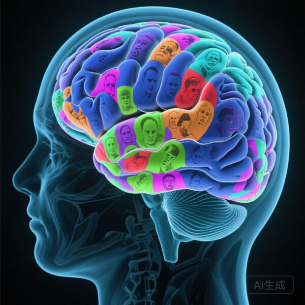
However, the normative modeling analysis told a fundamentally different story, exposing remarkable heterogeneity that group-level analysis concealed entirely. The central finding demonstrates that despite pervasive group differences, no more than 23% of TBI patients shared an extreme deviation in any single brain region. This means that in the vast majority of cases, an individual patient’s pattern of brain abnormalities bore little resemblance to other patients’ patterns—a striking illustration of how group averages can mislead clinical interpretation.
Perhaps most remarkably, while the group-level analysis suggested uniform brain changes across the TBI population, normative modeling revealed that every one of the 178 examined brain regions exhibited at least one extreme positive or negative deviation in at least one individual patient. This finding indicates that TBI can affect virtually any neuroanatomical structure in at least some patients, with the specific pattern of involvement varying enormously from person to person.
The study further examined whether injury severity, as measured by the Glasgow Coma Scale (GCS), might predict more consistent neuroanatomical involvement. Consistent with clinical intuition, the median number of extreme deviations increased progressively with injury severity: 9 deviations in patients with GCS 13-15 (mild injury), 19 in those with GCS 9-12 (moderate injury), and 22 in patients with GCS 3-8 (severe injury). This dose-response relationship validates the biological plausibility of the normative modeling approach while demonstrating that even severe injuries produce heterogeneous rather than uniform brain changes.
Critically, stratifying patients by injury severity did not eliminate the heterogeneity problem. Even within the most homogeneous subgroup examined (GCS 3-8), regional convergence did not exceed 34%, meaning that fewer than one in three severe TBI patients shared an extreme deviation in the same brain region. Even for patients with mild injuries typically considered to have favorable outcomes, the median of 9 extreme deviations indicates that subtle but potentially clinically meaningful neuroanatomical abnormalities occur in a highly individualized pattern.
Clinical Implications and Expert Commentary
These findings carry substantial implications for both research and clinical practice in traumatic brain injury. The study demonstrates that relying exclusively on group-level comparisons may provide an incomplete—and potentially misleading—picture of TBI-related brain changes. Normative modeling offers a complementary approach that respects the individuality of each patient’s brain injury while still enabling valid comparisons against expected ranges.
The concept of generating individualized “morphologic fingerprints” emerges as a particularly promising application of these methods. Just as clinical practice increasingly recognizes the value of personalized medicine approaches in oncology and other fields, neuroimaging may soon enable truly personalized assessment of brain injury consequences. Such approaches could potentially improve prognostic accuracy by identifying individual-specific risk profiles rather than relying on group-average trajectories.
The finding that heterogeneity persists even within narrow severity categories suggests that current classification systems based primarily on initial injury severity provide incomplete information about individual outcomes. Normative modeling might eventually supplement or refine these classification schemes by incorporating individual-specific deviation patterns that better capture the biological diversity of TBI pathophysiology.
Several limitations warrant consideration when interpreting these results. The study focused specifically on moderate-to-severe TBI populations, and findings may not generalize to milder injuries or to populations with different demographic characteristics than those represented in the ENIGMA Consortium cohort. Additionally, the cross-sectional design captures neuroanatomical status at a single timepoint, and longitudinal studies will be needed to determine how individual deviation patterns evolve over time and relate to functional outcomes.
The normative reference dataset itself may introduce limitations, as it represents a specific healthy population that may not perfectly match the demographic characteristics of all TBI patients. Methodological refinements in normative modeling approaches will likely address some of these concerns in future investigations.
Nevertheless, this study represents a significant methodological advance in TBI neuroimaging research. The integration of normative modeling into the analytical toolkit offers researchers and clinicians a more nuanced framework for understanding brain injury consequences that honors the fundamental complexity of this condition.
Conclusion: Toward Individualized Neuroimaging Assessment
The investigation by Mitchell and colleagues demonstrates that normative modeling reveals participant-specific cortical and subcortical abnormalities that conventional group comparisons overlook, more accurately representing the true diversity of TBI-related morphologic changes. By generating individualized deviation profiles rather than group averages, this approach enables detection of unique neuroanatomical signatures that may prove more informative for clinical decision-making than traditional categorical diagnoses.
The progressive increase in morphologic deviations with injury severity validates the biological relevance of these individual patterns while confirming that heterogeneity characterizes even the most severe injuries. Future research should explore how these individualized deviation profiles relate to long-term functional outcomes, cognitive recovery, and treatment response, potentially enabling more precise prognostication and personalized intervention strategies.
Normative modeling represents a paradigm shift in how clinicians and researchers conceptualize brain injury assessment, moving from population-based generalizations toward individual-specific characterization. As these methods continue to be refined and validated, they hold promise for advancing both scientific understanding and clinical care for the millions of individuals affected by traumatic brain injury worldwide.
Funding
Data used in this study were obtained from the ENIGMA Consortium Adult Moderate-to-Severe TBI working group. Supporting funding sources are detailed in the original publication.
References
1. Mitchell J, McDonald SJ, Law M, et al. Characterizing Heterogeneity in Brain Morphology in Traumatic Brain Injury Using Normative Modeling. Neurology. 2026;106(8):e214741. PMID: 41931749.
2. Bigler ED, Maxwell WL. Neuroimaging outcomes in traumatic brain injury. J Neurotrauma. 2022.
3. Marquand AF, Kia SM, Zabihi M, et al. Conceptualizing mental disorders as deviations from normative functioning. Mol Psychiatry. 2019.
4. ENIGMA Consortium. ENIGMA and the ENIGMA Transitional Injury Working Group protocols and documentation. 2023.