Introduction
Glaucoma is a progressive optic neuropathy characterized by structural damage to the retinal nerve fiber layer (RNFL) and corresponding visual field loss, often associated with elevated intraocular pressure (IOP). Clinicians typically rely on office IOP measurements to guide treatment and monitor disease control. However, despite apparently well-controlled office pressures, some patients continue to experience rapid functional and structural progression, highlighting a gap in risk stratification. This study evaluates the utility of an IOP stress test, specifically the water-drinking test (WDT), to identify glaucoma eyes at risk of faster disease progression beyond what routine office tonometry reveals.
Methods
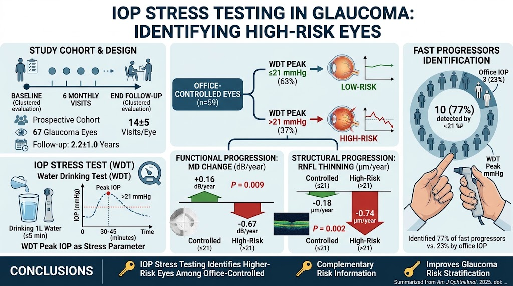
A prospective cohort study included 67 eyes from 36 glaucoma patients. Participants underwent the standardized WDT, an IOP stress test involving rapid water ingestion to provoke transient IOP spikes. Visual field (VF) tests and spectral-domain optical coherence tomography (SD-OCT) assessments of the RNFL were performed in clustered visits (five visits each) at baseline and study end, with intermediate single visits every six months, ensuring robust longitudinal monitoring. Rates of mean deviation (MD) change in visual fields and RNFL thinning were calculated using ordinary least-squares regression.
Associations between IOP parameters derived from the WDT—particularly peak IOP after water ingestion—and progression rates were analyzed. Analyses also considered severity-specific target office IOP thresholds to stratify eyes. Multivariable generalized estimating equation (GEE) models examined WDT peak IOP’s predictive value after adjusting for office IOP and other clinical factors, to assess whether the stress test provided independent prognostic information.
Results
The mean follow-up period after the WDT was 2.2 years, with an average of 14 testing visits per eye, enabling a detailed longitudinal assessment. Among 59 eyes with office IOP within severity-specific targets, 22 eyes (37%) exhibited WDT peak IOP values exceeding 21 mmHg. This subgroup showed significantly faster rates of visual field mean deviation loss (-0.67 dB/year compared to +0.16 dB/year, P=0.009) and RNFL thinning (-0.74 µm/year versus -0.18 µm/year, P=0.002) than eyes with controlled peak WDT IOP.
Thirteen eyes (19%) were classified as fast progressors based on their rates of functional and structural loss. Most of these eyes (77%) would have been assessed as controlled by office tonometry alone, with only 3 out of 13 exceeding target office IOP levels. However, 10 of these 13 fast progressors had WDT peak IOP above 21 mmHg, indicating the stress test’s sensitivity in unmasking hidden progression risk.
Multivariable GEE models demonstrated that peak WDT IOP >21 mmHg remained independently associated with accelerated MD loss (β=-0.659 dB/year, P=0.006) and RNFL thinning (β=-0.467 µm/year, P=0.035) after adjusting for office IOP and relevant covariates. Modeling WDT peak and office IOP as continuous variables yielded similar patterns, strengthening evidence of complementary prognostic value.
Discussion
This study supports the clinical utility of IOP stress testing, specifically via the WDT, to identify glaucoma patients at elevated risk of rapid progression despite apparently controlled office pressures. The WDT likely exposes transient IOP spikes that office measurements miss, which may contribute to ongoing optic nerve damage in susceptible eyes.
Identifying these high-risk eyes facilitates more personalized management—potentially prompting earlier or intensified treatment interventions to better preserve vision. The complementary information from the WDT provides a valuable adjunct to standard office tonometry, which alone may underestimate progression risk.
Furthermore, the WDT is a simple, noninvasive, low-cost test easily implementable in clinical practice. Its ability to predict structural and functional deterioration aligns with pathophysiologic understanding that IOP fluctuations and peaks contribute to glaucomatous damage beyond mean pressure levels.
Clinical Implications and Future Directions
Incorporating IOP stress testing into routine glaucoma care may refine risk stratification and guide treatment escalation decisions, especially for patients with controlled office IOP but unexplained progression. Clinicians should consider using the WDT or analogous stress tests for patients exhibiting suspicious progression or when office measurements fail to explain disease course.
Future research could expand on optimizing WDT protocols, exploring alternative stress tests, and integrating these with emerging biomarkers of glaucoma progression. Additionally, larger multicenter studies could validate the findings and establish standardized threshold values for risk categorization across diverse populations.
Conclusion
Intraocular pressure stress testing via the water-drinking test identifies glaucoma eyes at risk of faster structural and functional progression despite apparently controlled office pressures. This approach provides prognostic information complementary to routine office tonometry, supporting its role in enhanced glaucoma management to prevent vision loss. Clinicians are encouraged to consider incorporating IOP stress tests in patients with ongoing progression despite controlled office IOP to improve personalized care.
Reference
Malek DA, Medeiros FA. IOP Stress Testing Identifies Glaucoma Eyes at Risk of Fast Progression Despite Controlled Office Pressure. Am J Ophthalmol. 2026 Aug 12:S0002-9394(26)00454-X. doi: 10.1016/j.ajo.2026.08.012. Epub ahead of print. PMID: 42586188; PMCID: PMC13505876.