Introduction
Perforation of intrauterine devices (IUDs) is an uncommon yet significant complication occurring in approximately 0.2 to 3.6 per 1,000 insertions. This adverse event often necessitates surgical retrieval of the displaced device to prevent sequelae such as chronic pelvic pain, adhesions, or injury to intra-abdominal organs. Traditionally, removal of perforated IUDs has relied upon multiport laparoscopic techniques, commonly involving two or three incisions to allow triangulation of instruments and enhanced operative control. However, the continuous evolution of minimally invasive surgery has directed efforts towards minimizing surgical trauma, pain, and recovery time. This article delineates a pioneering approach utilizing a hysteroscope inserted transabdominally via a single laparoscopic port for perforated IUD extraction, illustrating the technique’s feasibility and clinical applicability.
Clinical Context and Disease Burden
IUDs are widely utilized for long-term reversible contraception given their efficacy and safety profile. Nonetheless, device perforation disrupts this safety, imposing morbidity and resource utilization burdens. The clinical challenge lies in safe and effective removal while mitigating operative risks. Conventional laparoscopy requires multiple ports to accommodate rigid laparoscopes and graspers, potentially increasing postoperative pain and risk of port-site complications. Hence, a single-port technique utilizing a flexible hysteroscope, primarily designed for intrauterine visualization, represents a valuable innovation for accessing and retrieving intra-abdominal foreign bodies.
Technical Overview
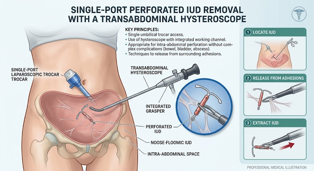
The described technique involves deploying a hysteroscope along with a specialized hysteroscopic grasper through one laparoscopic trocar introduced transabdominally. This single-port setup reduces incisional trauma and procedural complexity. The hysteroscopic instruments afford enhanced maneuverability within the abdominal cavity, facilitating precise localization, atraumatic adhesion dissection, and secure extraction of the perforated device. This approach is particularly suitable for patients without concomitant complications such as bowel or bladder injury or abscess formation, where more extensive surgical intervention might be warranted.
Key Findings and Clinical Implications
Clinical experience shared by Marguerie et al. demonstrates that despite varying challenges posed by differing IUD types and adhesion severity, the single-port transabdominal hysteroscopic method is effective in safely retrieving perforated devices. The versatility of the hysteroscopic grasper allows delicate manipulation minimizing collateral tissue damage. Moreover, the single-port approach streamlines the operation and can be expanded by adding additional ports if visual or instrument access is inadequate. This innovation aligns with the overarching goals of minimally invasive gynecologic surgery—maximal efficacy with minimal patient morbidity.
Expert Commentary and Perspectives
This technique represents a prime example of cross-applying existing gynecologic instrumentation to new surgical challenges. The use of a hysteroscope transabdominally reflects a creative adaptation improving operative ergonomics. Limitations include potential difficulty in complex adhesiolysis beyond the capabilities of hysteroscopic instruments alone and the necessity of carefully selecting candidates without organ injury or infection. Experts emphasize the importance of surgeon training and cautious intraoperative decision-making to transition seamlessly to multiport laparoscopy if indicated. Further studies comparing clinical outcomes and cost-effectiveness of this single-port approach versus traditional methods could solidify its role in clinical practice.
Conclusion and Future Directions
The single-port transabdominal hysteroscopic technique offers a promising minimally invasive option for perforated IUD removal, reducing operative trauma and enhancing patient recovery. Adoption of this approach should be considered in suitable patients, with readiness to escalate surgical access as clinically dictated. Continued innovation, training, and outcome reporting will be essential to optimize this technique’s safe integration into gynecologic surgery. Future research may also explore technological refinements such as dedicated hysteroscopic instruments optimized for intra-abdominal use and long-term follow-up assessing patient-centered outcomes.
Funding and Clinical Trials
No specific funding or clinical trial registration was reported in association with this technique.
References
1. Marguerie M, Andrew L, Sanders AP. Single-Port Perforated Intrauterine Device Removal With a Transabdominal Hysteroscope. Obstet Gynecol. 2026;148(3):e186–e188. doi:10.1097/AOG.0000000000004836
2. Kaislasuo J, Heikinheimo O, Lähteenmäki P. Incidence and risk factors for uterine perforation among 29, 898 insertions of intrauterine devices: a population-based study. Hum Reprod. 2013;28(5): 1248–1255.
3. Ahmad G, Ross JL, Shaktawat C, et al. Laparoscopic removal of migrated intrauterine contraceptive devices: a systematic review. Int J Gynaecol Obstet. 2010;108(3): 193–198.