Innovations in Esthetic Zone Implantation: Digital Integration of the Socket-Shield Technique with Guided Immediate Implant Placement and One-Step Crown
The esthetic zone in dental implantology, typically involving the anterior maxillary teeth, presents unique challenges due to its high demand for functional durability and esthetic perfection. Tooth extraction in this area often leads to alveolar bone loss and soft tissue remodeling, compromising implant success and esthetic outcomes. The socket-shield technique (SST) has been proposed as a conservative approach to preserve the buccal root fragment along with its periodontal attachment to minimize post-extraction bone resorption. Despite its advantages, the technique remains technically demanding, especially regarding accurate root fragment preparation.
Recent advances in digital dentistry, including computer-aided design/computer-aided manufacturing (CAD/CAM) and cone beam computed tomography (CBCT), have facilitated more precise surgical workflows. Integrating SST with fully digital guided implant placement may enhance procedural accuracy and reproducibility.
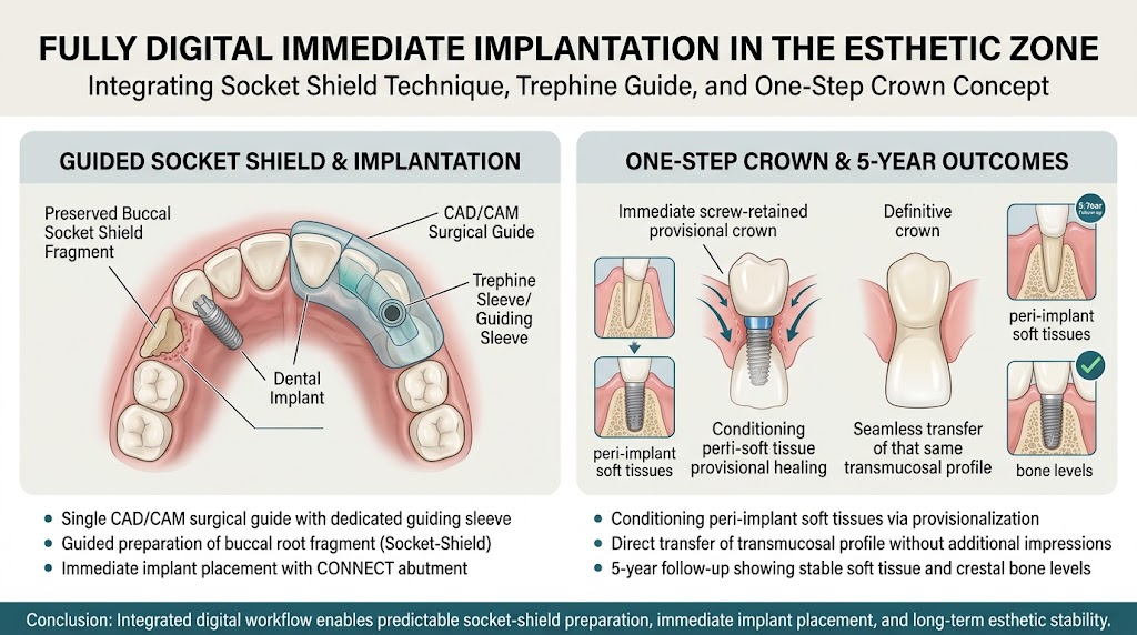
This article critically examines a recently published protocol involving a fully digital immediate implantation in the esthetic zone using the socket-shield technique combined with a novel trephine guide and the one-step crown concept, based on a 5-year clinical follow-up case.
Study Background
The conventional implant placement after tooth extraction in the esthetic zone often results in dimensional changes of the alveolar ridge and soft tissue recession, which pose esthetic challenges. The SST attempts to mitigate these changes by preserving the buccal root fragment to maintain the bundle bone and periodontal ligament complex, thus reducing post-extraction bone remodeling.
Nonetheless, the SST is intricate, primarily due to the difficulty in preparing the buccal root fragment while avoiding damage to the implant bed or adjacent anatomical structures. Inefficient root preparation may compromise treatment outcomes.
Digital workflows using CBCT and intraoral scanning now allow precise preoperative visualization and planning. The integration of CAD/CAM surgical guides can improve surgical accuracy, yet reports on combining these with SST are sparse.
Study Design and Methods
This investigation is centered on a single clinical case involving a 45-year-old female patient with a subgingival fracture of the maxillary right central incisor that contraindicated conservative or prosthetic restoration.
The protocol commenced with digital planning based on the superimposition of CBCT and intraoral scan data to precisely map the tooth root anatomy and adjacent bone.
A bespoke CAD/CAM surgical guide was fabricated incorporating a dedicated guiding sleeve designed for trephine-like controlled root fragment preparation. This guide served a dual function:
1. Guiding the atraumatic preparation of the buccal root fragment (socket shield).
2. Facilitating immediate implant placement in an ideal three-dimensional position.
After socket-shield preparation and implant placement, a transmucosal CONNECT abutment was installed. Immediate provisionalization was performed using a crown fabricated to condition the peri-implant soft tissues.
Notably, the provisional restoration contour was transferred unchanged to the definitive crown using a one-step crown concept, obviating the need for additional intraoral scans or conventional impressions, thereby streamlining the prosthetic workflow.
Key Findings and Results
The patient was followed clinically and radiographically for five years post-procedure. Key outcomes included:
– Stable peri-implant soft tissue levels with no signs of recession or inflammation.
– Maintaining marginal bone levels around the implant without radiographic evidence of resorption or infection.
– No peri-implant complications such as infection, implant mobility, or prosthetic failures.
– Preservation of the buccal cortical plate contributing to natural esthetic gingival contours.
This case underscores that the digital integration of SST with guided implant placement and immediate provisionalization is feasible and may enhance the precision of root fragment preparation and implant positioning.
Expert Commentary
The technique-sensitive nature of SST has limited its routine application. The introduction of a dedicated guiding sleeve within a digital surgical guide represents a meaningful advancement, potentially mitigating operator variability and technical errors. By incorporating full digital datasets combining CBCT and intraoral scanning, the method allows comprehensive preoperative planning of root fragment dimensions and implant angulation.
However, it is paramount to recognize that this report is based on a single case study. Although the long-term follow-up is encouraging, larger prospective, randomized studies are essential to validate the accuracy, reproducibility, and clinical advantages over conventional protocols or non-guided SST.
Furthermore, translational challenges such as learning curve, equipment costs, and integration into everyday clinical workflows merit consideration. The one-step crown concept employed also requires further evaluation to confirm its generalizability and impact on esthetic outcomes across a broader patient population.
Conclusion
The presented fully digital protocol combining the socket-shield technique with a novel trephine guide and immediate implant placement in the esthetic zone demonstrates promising clinical feasibility and favorable 5-year outcomes in maintaining peri-implant tissue stability.
This approach potentially offers improved precision in root fragment preparation and implant positioning, enhancing esthetic results while streamlining the surgical and prosthetic workflows.
Nonetheless, further rigorous clinical trials are indispensable to substantiate these findings, optimize the technique, and assess its broader applicability.

 By
No Comments