Patient Information
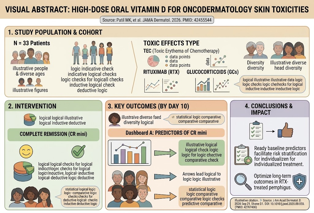
This retrospective multicenter case series included 33 patients (mean age 60.9 years, SD 14.6; 19 females, 58%) who developed toxic erythema of chemotherapy (TEC) or acute radiation dermatitis (ARD). Patients were treated across three academic medical centers between December 2021 and January 2024. Inclusion criteria mandated patients to have received one or two doses of high-dose oral vitamin D (100,000 international units [IU]) with at least 10 days of clinical follow-up.
The majority suffered from TEC (28 patients, 85%), with the remainder presenting with ARD (5 patients, 15%). All patients manifested severe cutaneous toxic effects, characterized primarily by erythema and symptomatic discomfort such as pain and burning sensations, frequently necessitating interruptions or modifications of their ongoing chemotherapy or radiation therapy regimens.
Diagnosis
Diagnosis was established based on clinical features consistent with toxic erythema of chemotherapy or acute radiation dermatitis. TEC presents as erythematous patches, plaques, or bullous lesions often triggered by cytotoxic agents, with histologic findings occasionally indicative of neutrophilic eccrine hidradenitis or Stevens-Johnson syndrome/toxic epidermal necrolysis (SJS/TEN)-like patterns, which were confirmed or suspected in some subtypes.
ARD diagnosis relied on the temporal association with radiation treatment sites, showing erythema and skin barrier disruption due to acute radiation exposure. The clinical diagnosis was supported by detailed medical histories, physical examinations, and exclusion of infectious or other inflammatory etiologies.
Differential Diagnosis
The differential diagnoses considered included:
– Infectious cellulitis or erysipelas: Ruled out by absence of systemic signs of infection and rapid response to immunomodulatory treatment without antibiotics.
– Allergic contact dermatitis: Less likely due to lack of exposure history and distribution patterns.
– Drug hypersensitivity reactions not related to oncologic treatments: Excluded by temporal relationship to chemotherapy or radiation.
– Autoimmune blistering diseases: Unlikely due to clinical presentation and rapid response to vitamin D treatment.
These alternate diagnoses were excluded based on clinical criteria, laboratory data where applicable, and thorough patient evaluations.
Treatment and Management
Patients received one or two oral doses of high-dose vitamin D (cholecalciferol or ergocalciferol) at 100,000 IU per dose. The treatment approach was initiated with the rationale that vitamin D acts as a rapid immunomodulator, potentially mitigating inflammatory skin damage caused by chemotherapy and radiation.
The dosing was administered under medical supervision with monitoring of serum calcium levels to detect hypercalcemia risk. Supportive care, including symptomatic measures such as emollients and analgesics, was provided as necessary. The intervention aimed to reduce cutaneous inflammation rapidly to allow continuation of anticancer therapies without interruption.
Outcome and Prognosis
Efficacy outcomes included both patient-reported symptomatic relief and objective clinician assessment of erythema using a 5-point Likert scale. Among 30 patients with reported follow-up data, 26 (87%) experienced symptomatic relief within 10 days of treatment; median time to improvement was 5 days (range 1-28), notably faster (median 3 days) for inpatients.
Clinician-assessed erythema severity decreased from a mean Likert score of 4.36 at baseline to 2.21 by day 10, indicating significant improvement. Patients with neutrophilic eccrine hidradenitis and SJS/TEN-like TEC subtypes demonstrated particularly rapid resolution.
No significant alterations in serum calcium were observed, and importantly, 24 of the 33 patients (73%) could continue anticancer therapy without interruption, underscoring the clinical utility of this intervention. No treatment-related adverse events were reported during the study period.
Discussion
Severe cutaneous toxic effects related to chemotherapy and radiation frequently cause treatment delays, dose reductions, and impact quality of life adversely. Current management strategies often rely on topical steroids and symptomatic care, with limited options for rapid systemic modulation of the inflammatory response.
This case series is among the first to document the use of high-dose oral vitamin D as a potentially effective and safe immunomodulatory agent in skin toxicity secondary to cancer treatments. Vitamin D’s role in immune regulation, inflammation attenuation, and skin barrier maintenance is well-documented in preclinical models and select human conditions. However, robust clinical data for its application in oncodermatology have been scarce.
The findings here reveal that a single or repeated high oral dose of vitamin D can lead to rapid clinical improvement, symptom relief, and enable continued anticancer treatment. The safety profile with no adverse effects and the absence of hypercalcemia are reassuring, although longer-term studies are warranted.
Notably, subtypes associated with intense neutrophilic infiltration (neutrophilic eccrine hidradenitis) and severe epidermal damage (SJS/TEN-like patterns) exhibited quick responses, suggesting a particular benefit in these presentations. Mechanistically, vitamin D may inhibit proinflammatory cytokine cascades, modulate epidermal keratinocyte behavior, and promote regeneration.
Limitations include the retrospective design, small sample size, and lack of a control group. Despite these, the multicenter nature enhances generalizability. Prospective randomized controlled trials are needed to confirm efficacy, optimal dosing, and long-term safety.
References
1. Patil MK, Sakunchotpanit G, Toulmin SA, et al. High-Dose Oral Vitamin D for Toxic Effects of the Skin Associated With Chemotherapy and Radiation. JAMA Dermatol. 2026 Sep 1;162(9):888-893. doi:10.1001/jamadermatol.2026.5554. PMID: 42455544.
2. Lewiecki EM, et al. The role of vitamin D in the skin: recent advances and perspectives. Dermatoendocrinology. 2011 Jan 1;3(4):240-241.
3. Blakely KM, et al. Management of cutaneous side effects of chemotherapy and radiotherapy. J Support Oncol. 2006 Sep-Oct;4(7):353-358.
4. Bikle DD. Vitamin D and the skin: physiology and pathophysiology. Rev Endocr Metab Disord. 2012 Mar;13(1):3-19.
—
This case series highlights an emerging supportive care strategy addressing a significant clinical problem in oncology and dermatology, combining clinical observation with immunomodulatory pharmacology for improved patient outcomes.