Highlight
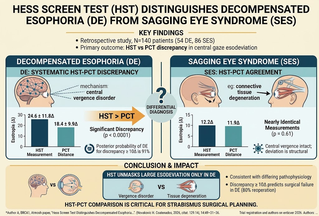
– The Hess Screen Test (HST) reveals significantly larger esodeviation in patients with decompensated esophoria (DE) compared to measurements by prism cover test (PCT).
– In contrast, patients with sagging eye syndrome (SES) show minimal discrepancy between HST and PCT measurements.
– An HST-PCT discrepancy of ≥10 prism diopters (Δ) predicts a high likelihood (91% posterior probability) of DE.
– Persistent discrepancy postoperatively in DE patients correlates with a higher reoperation rate, emphasizing its prognostic value.
Study Background
Acquired esotropia represents a substantial clinical challenge, particularly in distinguishing between decompensated esophoria (DE) and esotropia caused by sagging eye syndrome (SES). DE is primarily a central vergence disorder leading to a failure in maintaining binocular alignment, whereas SES is a mechanical disorder resulting from connective tissue degeneration affecting orbital support. Accurate diagnosis is essential, as treatments and prognostic implications differ importantly between these entities. However, conventional clinical assessments like the prism cover test (PCT) may lack sensitivity in differentiating these conditions. This study evaluates the diagnostic performance of the Hess Screen Test (HST), a subjective ocular motility assessment, in distinguishing DE from SES and guiding surgical decision-making.
Study Design
This retrospective comparative cross-sectional and cohort study included 140 patients presenting with acquired esotropia due to either DE (n=54) or SES (n=86). Inclusion criteria targeted adult patients with documented esodeviation assessed clinically. The primary endpoint was the difference in central gaze esotropia measured by HST versus PCT at both distance and near fixation. The study further examined the posterior probability of DE diagnosis based on the magnitude of HST-PCT discrepancy using Bayesian analysis. For patients undergoing surgical intervention, postoperative persistence of the discrepancy was evaluated as a predictor for the necessity of reoperation.
Key Findings
The mean esotropia measured by HST in DE patients was 24.6±11.8Δ, significantly exceeding the PCT measurement at distance fixation (18.4±9.9Δ, p<0.0001). Conversely, SES patients showed near-identical measurements using both techniques (12.2Δ by HST vs. 11.9Δ by PCT; p=0.61). This distinct pattern reflects the different pathophysiologic mechanisms: DE involves central vergence control dysfunction causing variable and often greater esodeviation under specific testing conditions, whereas SES-related esotropia stems from static connective tissue laxity and orbital mechanical changes, resulting in consistent measurements between tests.
A threshold discrepancy of 10Δ or greater between HST and PCT measurements yielded a 91% posterior probability for DE diagnosis, demonstrating strong diagnostic utility. Importantly, among DE patients treated surgically, those with persistent postoperative HST-PCT discrepancy ≥10Δ had an 80% reoperation rate, contrasted with only 14% in patients with lower discrepancy (p=0.008). This finding indicates that residual central vergence abnormalities, detectable by HST but masked on PCT, predict surgical failure or incomplete correction, thereby guiding follow-up and management.
Expert Commentary
The differentiation between DE and SES has been a clinical conundrum due to overlapping clinical features but fundamentally different pathophysiologic underpinnings. This study provides compelling evidence supporting the Hess Screen Test as a superior modality to uncover latent central vergence dysfunction in DE, which is not appreciated by standard prism cover testing. As Vergence is a complex neural mechanism influencing binocular alignment, tests that engage more dynamic ocular motor control, such as the HST, can unmask larger deviations that represent the true burden of disease in DE patients.
The study’s use of Bayesian posterior probability offers a quantitative approach to applying HST-PCT discrepancy thresholds, enhancing diagnostic confidence. Moreover, the prognostic implications regarding reoperation risk fills a critical gap in surgical management strategies for acquired esotropia. However, as a retrospective study, limitations include potential selection bias and variability in clinical testing conditions. Prospective validation and standardized testing protocols would strengthen generalizability.
Mechanistically, the findings align with current understanding: SES involves mechanical disruption of extraocular muscle pulleys and connective tissues leading to relatively fixed ocular misalignment, while DE reflects a failure of central vergence control, which is more variable and dynamic. This has implications for therapeutic goals, where DE may benefit from approaches targeting neural control (e.g., prism therapy, vision orthoptics) alongside surgery.
Conclusion
The Hess Screen Test emerges as a valuable diagnostic and prognostic tool that discriminates decompensated esophoria from sagging eye syndrome by revealing a hallmark discrepancy with prism cover test measurements in DE patients. This difference underscores their distinct pathophysiological basis—central vergence disorder versus connective tissue degeneration. Incorporating HST into routine evaluation may enhance diagnostic accuracy, tailor surgical planning, and identify patients at elevated risk for reoperation. Future prospective studies are warranted to validate thresholds and determine how best to integrate HST findings into clinical algorithms for acquired esotropia.
Funding and ClinicalTrials.gov
The study did not report specific funding sources. ClinicalTrials.gov registration details were not provided.
References
- Yehezkeli V, Parunakian E, Neimark E, Demer JL. Hess Screen Test Distinguishes Decompensated Esophoria From Esotropia Due To Sagging Eye Syndrome. American Journal of Ophthalmology. 2026; (published online 2026 Sep 23). PMID: 42777795.
- Coats DK, Paysse EA, Cuppers C. Sagging eye syndrome: clinical features and surgical management. J AAPOS. 2007;11(4):367-373.
- Demer JL. The Orbital Connective Tissue in Sagging Eye Syndrome. Invest Ophthalmol Vis Sci. 2016;57(12):5263-5273.
- Guyton DL. Decompensated Esophoria and Central Vergence Influences. Arch Ophthalmol. 1989;107(7):954-957.