Patient Information
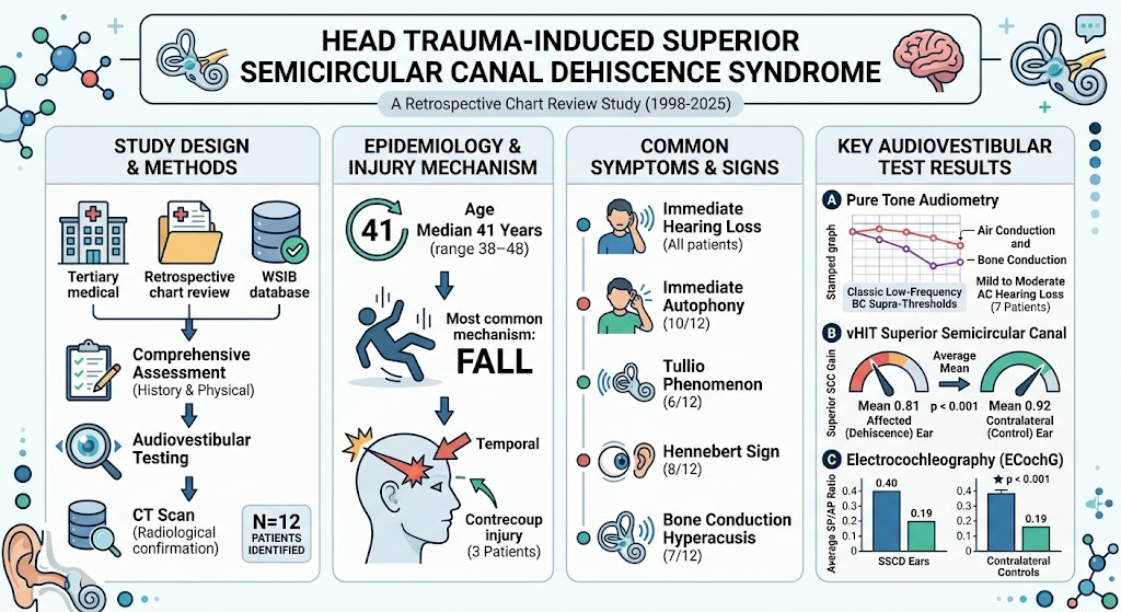
This case series includes twelve patients identified retrospectively from the Province of Ontario Workplace Safety and Insurance Board database spanning 1998 to 2025. The median age was 41 years (IQR 38-48), with all presenting following minor head trauma. The precipitating events were predominantly falls impacting the temporal region, with three patients exhibiting a contrecoup injury pattern. All patients reported immediate hearing loss after injury. Additional presenting symptoms included autophony in 10 patients, Tullio phenomenon (sound-induced vertigo) in six, Hennebert sign (pressure-induced nystagmus) in eight, and bone conduction hyperacusis in seven.
Diagnosis
All patients underwent comprehensive audiovestibular assessments alongside high-resolution computed tomography (CT) scans of the temporal bones to confirm superior semicircular canal dehiscence (SSCD). Pure tone audiometry revealed classical low-frequency bone conduction suprathresholds and mild to moderate air conduction hearing loss in seven patients. Video head impulse testing (vHIT) demonstrated significant unilateral reduction in superior semicircular canal gain (mean 0.81 vs. 0.92 contralaterally, p<0.001), indicative of a vestibular deficit localized to the affected canal. Electrocochleography showed elevated summating potential/action potential (SP/AP) ratios in SSCD ears (0.40) compared to contralateral ears (0.19), supporting the presence of a third mobile window effect.
The combination of immediate hearing loss after head trauma, audiological profiles suggestive of low-frequency air-bone gap, vestibular hypersensitivity signs, and confirmatory CT imaging established the diagnosis of trauma-induced SSCD syndrome.
Differential Diagnosis
Differential diagnoses considered included labyrinthine concussion, perilymphatic fistula, otosclerosis, vestibular migraine, and other causes of conductive hearing loss with vertigo. Labyrinthine concussion typically lacks radiological evidence of canal dehiscence and may present without autophony or Tullio phenomenon. Perilymphatic fistula was less likely due to absence of active fluid leak evidence. Otosclerosis usually demonstrates progressive conductive loss without overt vestibular symptoms. Vestibular migraine was excluded by acute onset post-trauma and audiovestibular test findings. The diagnostic imaging and electrophysiological data were essential to distinguish SSCD from these mimics.
Treatment and Management
Management was individualized based on symptom severity. Initial conservative approaches included vestibular rehabilitation and hearing support. For significant vestibular symptoms and disabling autophony, surgical repair via canal plugging or resurfacing was considered. Patients were counseled on the risk of progression and the nature of SSCD as a third window pathology. Audiology follow-up was arranged to monitor hearing status, and serial vestibular testing was employed to assess functional recovery or deterioration.
Outcome and Prognosis
Outcomes varied among patients; those with mild symptoms improved with conservative management over months. Surgical candidates reported symptomatic relief post-intervention, with reduction in vestibular hypersensitivity and improvement in quality of life. The prognosis is favorable if identified early, though residual vestibular deficits may persist. Recognition of minor head trauma as a trigger for SSCD syndrome underscores the importance of timely diagnosis and management to prevent long-term morbidity.
Discussion
Superior semicircular canal dehiscence syndrome is classically described in patients with congenital or idiopathic thinning or absence of the bony roof overlying the superior semicircular canal, producing a “third window” effect that alters cochlear and vestibular function. While radiologic SSCD has been identified in asymptomatic populations, this series emphasizes how seemingly minor head trauma can precipitate a clinically significant syndrome in anatomically predisposed individuals.
The findings underscore the importance of comprehensive vestibular and audiological testing in patients presenting with sudden hearing loss and vestibular symptoms after head injury. The vHIT’s ability to detect canal-specific gain reduction and elevated SP/AP electrocochleography ratios serve as important adjuncts in diagnosis. The predominance of temporal bone impact and occasional contrecoup injuries suggest biomechanical forces contribute to dehiscence development or symptom manifestation.
From a clinical standpoint, this awareness facilitates early recognition and treatment. Moreover, understanding that mild trauma can unmask SSCD may influence workplace safety considerations and medico-legal evaluations. Further prospective studies could delineate risk factors for progression and refine management algorithms.
References
1. Ungar OJ, O’Shea R, Rutka JA. Head Trauma-Induced Superior Semicircular Canal Dehiscence Syndrome. The Laryngoscope. 2026 Aug 27. doi:10.1002/lary.30001.
2. Minor LB, Solomon D, Zinreich JS, Zee DS. Sound- and pressure-induced vertigo due to bone dehiscence of the superior semicircular canal. New England Journal of Medicine. 1998 Oct 15;339(12):985-91.
3. Carey JP, Minor LB, Nager GT. Dehiscence or thinning of bone overlying the superior semicircular canal in patients with imbalance and pressure-induced vertigo. Archives of Otolaryngology–Head & Neck Surgery. 2000 Mar;126(3):334-40.
4. Rosowski JJ. Sensory hearing loss, hyperacusis, and noise-induced hearing loss. Otolaryngologic Clinics of North America. 2018 Feb;51(1):81-93.