Highlights
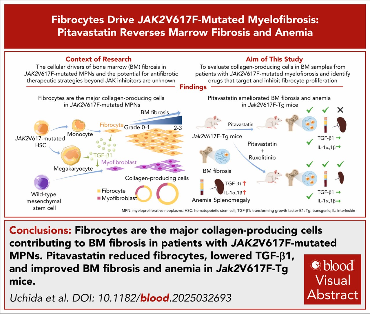
- Identification of fibrocytes as the predominant collagen-producing cells driving bone marrow fibrosis in JAK2V617F-mutated myelofibrosis.
- Pitavastatin demonstrated efficacy in reducing fibrocyte proliferation and bone marrow fibrosis in Jak2V617F murine models.
- Reduction of TGF-β1 production by neoplastic fibrocytes with pitavastatin leads to diminished myofibroblast expansion, linking hematopoietic fibrosis drivers to mesenchymal responses.
- Peripheral blood fibrocytes from patients with JAK2V617F-mutated MPNs also show sensitivity to pitavastatin, supporting translational potential.
Background
Myelofibrosis (MF) is a myeloproliferative neoplasm characterized by progressive bone marrow (BM) fibrosis, extramedullary hematopoiesis, cytopenias, and constitutional symptoms. The JAK2V617F mutation is a prevalent driver in primary and secondary MF, contributing to myeloproliferation and fibrosis. Traditionally, BM fibrosis has been attributed to a reactive, cytokine-mediated transformation of mesenchymal stromal cells (MSCs) into myofibroblasts, largely driven by TGF-β1 secreted by abnormal megakaryocytes and platelets. However, accumulating evidence suggests that hematopoietic-derived fibrocytes—monocyte-lineage cells producing extracellular matrix proteins—may be central to fibrogenesis in MF.
Despite therapeutic advances with JAK inhibitors, reversal of fibrosis and amelioration of anemia remain challenging, underscoring a need for novel antifibrotic strategies directly targeting fibrosis-driving cells. This review synthesizes the current state of knowledge concerning the role of fibrocytes in JAK2V617F-mutated MF and the emerging preclinical evidence supporting pitavastatin as a potential antifibrotic agent.
Key Content
1. Cellular and Molecular Pathogenesis of Bone Marrow Fibrosis in JAK2V617F-Mutated MF
The classical paradigm implicates cytokine-induced MSC differentiation into collagen- and fibronectin-producing myofibroblasts, catalyzed by TGF-β1 and other fibrogenic mediators primarily from mutated megakaryocytes. However, emerging work has characterized fibrocytes—monocyte-derived, hematopoietic lineage cells expressing markers such as CD45 and collagen—as significant contributors to extracellular matrix deposition in MF.
The study by Uchida et al. (Blood, 2026) quantitatively demonstrated that fibrocytes comprise approximately two-thirds of collagen-producing cells in BM biopsies from patients with JAK2V617F-mutated MPNs, surpassing myofibroblast contributions. These findings reshape the cellular framework of BM fibrosis, highlighting fibrocytes’ dominant role.
2. Experimental Models and Drug Screening for Fibrocyte Targeting Agents
Utilizing Jak2V617F transgenic mice, which recapitulate human MF pathology, bone marrow-derived fibrocytes were isolated and subjected to a high-throughput drug screen. Statins, particularly pitavastatin, surfaced as potent inhibitors of fibrocyte proliferation in vitro, marking a pioneering therapeutic candidate addressing fibrogenic hematopoietic cells.
This high-throughput approach represents a methodological advancement for identifying antifibrotic agents with cellular specificity beyond cytokine modulation.
3. Pitavastatin Efficacy In Vivo and Mechanistic Insights
In vivo pitavastatin administration in Jak2V617F mice led to a marked reduction in fibrocyte counts and bone marrow fibrosis severity, evaluated through histopathological grading and collagen quantification. Concurrently, improvements in anemia parameters were observed, indicating functional hematopoietic recovery.
Mechanistically, pitavastatin reduced TGF-β1 production from neoplastic fibrocytes, disrupting the paracrine signal driving myofibroblast differentiation and expansion, thereby attenuating fibrosis from both hematopoietic and stromal compartments. These findings suggest a dual antifibrotic action of pitavastatin targeting upstream fibrocytes and downstream stromal myofibroblasts.
4. Translational Evidence: Human Fibrocytes and Pitavastatin Sensitivity
Peripheral blood-derived fibrocytes isolated from patients harboring JAK2V617F mutations similarly showed significant sensitivity to pitavastatin-induced proliferation inhibition in vitro. This translational evidence supports potential clinical application and justifies further clinical trials to evaluate pharmacodynamic effects, safety, and efficacy in human MF.
5. Comparative Context and Emerging Therapeutic Landscape
Current antifibrotic treatments in MF remain limited. JAK inhibitors (e.g., ruxolitinib) mitigate symptom burden and splenomegaly but have limited impact on reversing fibrosis or correcting anemia. Agents targeting TGF-β signaling have been explored but lack clinical viability due to systemic toxicities.
By directly targeting fibrogenic fibrocytes and their cytokine output, pitavastatin offers a novel mechanism-based therapy with a favorable safety profile established from cardiovascular indications. The repositioning of statins in MF therapy aligns with trends in drug repurposing, enabling expedited translational steps.
Expert Commentary
The identification of fibrocytes as key effectors in JAK2V617F-mutated MF represents a paradigm shift from the MSC/myofibroblast-centric model to a hematopoietic cell-driven fibrosis framework. This underscores the heterogeneity and complexity of fibrogenesis in the BM microenvironment.
The translational potential of pitavastatin is compelling, given its dual antifibrotic and hematologic benefits demonstrated preclinically. However, despite promising murine data, clinical trials are essential to evaluate dose optimization, patient stratification by molecular mutation status, and long-term outcomes.
Potential limitations include the need to ascertain pitavastatin’s effects on non-fibrotic hematopoietic and stromal cell functions, potential off-target effects, and synergy or antagonism with established therapies like JAK inhibitors. The interplay between fibrocytes and myofibroblasts, especially through TGF-β1 signaling, illuminates complex cellular crosstalk that may require combination therapeutic approaches.
Furthermore, the study enhances understanding of pathophysiologic mechanisms linking mutated hematopoietic clones to extracellular matrix remodeling, providing a foundation for biomarker development and precision medicine in MF.
Conclusion
Recent evidence firmly establishes fibrocytes as principal contributors to bone marrow fibrosis in JAK2V617F-mutated myelofibrosis, challenging prevailing fibrogenic paradigms. Pitavastatin emerges as a promising repurposed agent capable of inhibiting fibrocyte proliferation, reducing TGF-β1-mediated myofibroblast expansion, and ameliorating marrow fibrosis and anemia in preclinical models.
These findings demand further clinical evaluation of pitavastatin for MF patients harboring JAK2V617F mutations, potentially expanding therapeutic options beyond symptom palliation to disease modification. Future research should focus on large-scale clinical trials, elucidation of molecular mechanisms, and integration within multimodal treatment regimens.
References
- Uchida T, Shide K, Kameda T, Ozono Y, Kubuki Y, Tahira Y, Kamiunten A, Marutsuka K, Akizuki K, Karasawa M, Uehira Y, Ueno H, Yamaguchi H, Shimoda K. Fibrocytes drive JAK2V617F-mutated myelofibrosis: pitavastatin reverses marrow fibrosis and anemia. Blood. 2026;148(13):1723-1736. PMID: 42391645.
- Kleppe M, Kwak M, Koppikar P, et al. JAK-STAT pathway activation in myeloproliferative neoplasms and potential therapeutic targeting. Hematol Oncol Clin North Am. 2018;32(2):203-217. PMID: 29305921.
- Steensma DP. Myelofibrosis biology and treatment. Hematology Am Soc Hematol Educ Program. 2021;2021(1):254-263. PMID: 34916506.
- Verstovsek S. Ruxolitinib in myelofibrosis: latest evidence and clinical potential. Ther Adv Hematol. 2015;6(4):196-206. PMID: 26120468.
- Mehrling T, Nolte F, Stegelmann F, et al. Fibrocytes as novel effectors in fibrotic diseases: implications in cancer and pulmonary disease. Front Med (Lausanne). 2020;7:569032. PMID: 33056645.