Background
Shock is a complex clinical syndrome characterized by insufficient tissue perfusion leading to cellular and organ dysfunction. Accurate and rapid identification of the etiology of shock in critically ill mechanically ventilated patients is vital to guide targeted therapeutic interventions and improve outcomes. Resuscitative transesophageal echocardiography (TEE) has emerged as a valuable bedside tool for hemodynamic monitoring and assessment of cardiac function in this setting. However, pulmonary contributions to shock states, including fluid overload, pneumothorax, consolidation, and pleural effusions, often require additional imaging for full elucidation.
Transesophageal lung ultrasound (TELUS) integrated alongside TEE is a novel approach that allows for simultaneous cardiac and pulmonary evaluation without repositioning the patient, potentially expediting diagnosis and management. Despite its theoretical advantages, systematic data regarding the feasibility, clinical utility, and impact of TELUS as an adjunct to resuscitative TEE in shock assessment remain limited.
Study Design
This multicenter observational cohort study analyzed prospectively collected data from the Resuscitative Transesophageal Echocardiography Collaborative Registry (rTEECoRe), involving 23 hospitals. The study focused on adult patients who were mechanically ventilated and underwent TEE for evaluation of shock or hemodynamic monitoring.
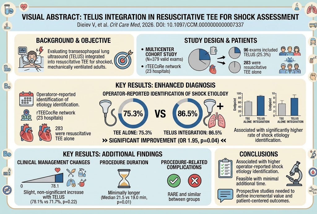
Out of 1213 TEE examinations documented in the registry, 379 met inclusion criteria specifically involving shock assessment or hemodynamic monitoring in mechanically ventilated patients. Within this subset, 96 examinations (25.3%) incorporated TELUS as an adjunct procedure. No additional interventions were applied beyond routine clinical care.
Primary endpoints included the feasibility of TELUS integration, operator-reported identification of shock etiology, and impact on clinical management decisions. Secondary analyses examined procedure duration, operator specialty performing TELUS, pulmonary ultrasound findings, and occurrence of procedure-related complications.
Key Findings
Operators reported a statistically significant increase in identification of shock etiology when TELUS was added to resuscitative TEE compared with TEE alone—86.5% versus 75.3% respectively (odds ratio [OR] 1.95; 95% confidence interval [CI] 1.02-3.72; p = 0.04). This suggests TELUS may enhance diagnostic clarity by incorporating lung imaging findings relevant to shock mechanisms.
Changes in clinical management were noted more frequently in the TELUS group (78.1%) versus the TEE-only group (71.7%), although this difference did not reach statistical significance (OR 1.40; 95% CI 0.82-2.41; p = 0.22). This trend indicates potential increased influence on treatment decisions but warrants further investigation.
TELUS procedures took slightly longer, with a median duration of 21.5 minutes compared to 19.0 minutes for TEE alone (p = 0.01), reflecting the additional imaging component. The majority of TELUS exams were performed by intensivists, highlighting the skillset and workflow integration within critical care teams.
Pulmonary findings detected by TELUS varied, including B-lines (indicative of interstitial syndrome or pulmonary edema), pleural effusions, lung consolidations, and A-line patterns (suggestive of normal aeration). These findings occasionally guided specific respiratory or antimicrobial interventions, illustrating clinical relevance in complex shock cases.
Importantly, procedure-related complications were uncommon and occurred at similar rates between TELUS and TEE groups, supporting the safety of this combined approach.
Expert Commentary
The integration of TELUS into resuscitative TEE is a promising advancement that leverages the anatomical proximity of the esophagus to assess thoracic structures efficiently in critically ill ventilated patients. By providing dynamic lung assessment alongside cardiac imaging, clinicians can detect concurrent pulmonary pathology that might otherwise be missed or delayed, facilitating more comprehensive shock evaluations.
Despite encouraging findings, the observational design and operator-reported outcomes highlight limitations including potential bias and lack of standardized TELUS acquisition and interpretation protocols. Additionally, study findings may be influenced by varying operator expertise and institutional protocols.
Future prospective, controlled studies with blinded independent adjudication are necessary to validate these results and to determine whether TELUS incorporation translates into improved patient-centered outcomes such as mortality, ventilator days, or ICU length of stay.
Conclusion
This multicenter observational study suggests that adding transesophageal lung ultrasound to routine resuscitative transesophageal echocardiography is feasible, safe, and associated with improved operator-reported identification of shock etiology in mechanically ventilated adults. Although changes in clinical management were not significantly different, trends favor TELUS utility. The modest increase in procedure duration is acceptable given the potential diagnostic benefits.
Standardization of TELUS protocols, training, and further robust clinical trials are needed to delineate the precise role and incremental value of TELUS in shock evaluation and management paradigms. Integration of combined cardiac and pulmonary ultrasound may represent an important evolution in critical care diagnostic modalities.
Funding and Registration
The study utilized registry data from the rTEECoRe network supported by participating centers. There were no specific interventions or additional funding disclosures reported. Clinical trial registration details were not indicated.
References
1. Dieiev V, Salinas PD, Teran F, et al. Feasibility and Operator-Reported Utility of Transesophageal Lung Ultrasound As an Adjunct to Resuscitative Transesophageal Echocardiography in the Evaluation of Shock: A Multicenter Observational Study. Crit Care Med. 2026 Sep 4. PMID: 42695753.
2. Vieillard-Baron A, Price S, et al. Echocardiography in the intensive care unit: from evolution to revolution? Intensive Care Med. 2018;44(5):747-749.
3. Lichtenstein DA. Lung ultrasound in the critically ill. Ann Intensive Care. 2014;4(1):1.
4. Vieillard-Baron A. Monitoring volume status and fluid responsiveness by echocardiography. Anaesth Crit Care Pain Med. 2019;38(6):659-664.
5. Volpicelli G, Elbarbary M, et al. International evidence-based recommendations for point-of-care lung ultrasound. Intensive Care Med. 2012;38(4):577-591.