Highlight
This study identifies an optimized low-dose CT scan emphysema threshold that moderately detects previously undiagnosed COPD among lung cancer screening participants. Incorporating quantitative airway metrics such as airway wall thickness and airway branch count with clinical characteristics significantly improves detection accuracy while reducing unnecessary spirometry referrals. A machine-learning ensemble model (Extreme Gradient Boosting) integrating these parameters outperformed emphysema assessment alone in identifying airflow obstruction in current and former smokers.
Study Background
Chronic obstructive pulmonary disease (COPD) and lung cancer share a common risk factor: tobacco smoke exposure. Lung cancer screening programs employing low-dose computed tomography (CT) present a unique opportunity to opportunistically detect undiagnosed COPD in high-risk individuals. Early identification of COPD is critical to timely intervention, potentially improving outcomes and reducing healthcare burden. However, guidelines on when to refer lung cancer screening patients for confirmatory spirometry based on CT findings are limited. Traditional COPD diagnosis relies on spirometry to detect fixed airflow obstruction, but widespread spirometry screening is challenging in clinical practice. Quantitative CT biomarkers, including emphysema extent and airway morphology, increasingly provide structural insights that may augment clinical assessment and predict airflow obstruction.
Study Design
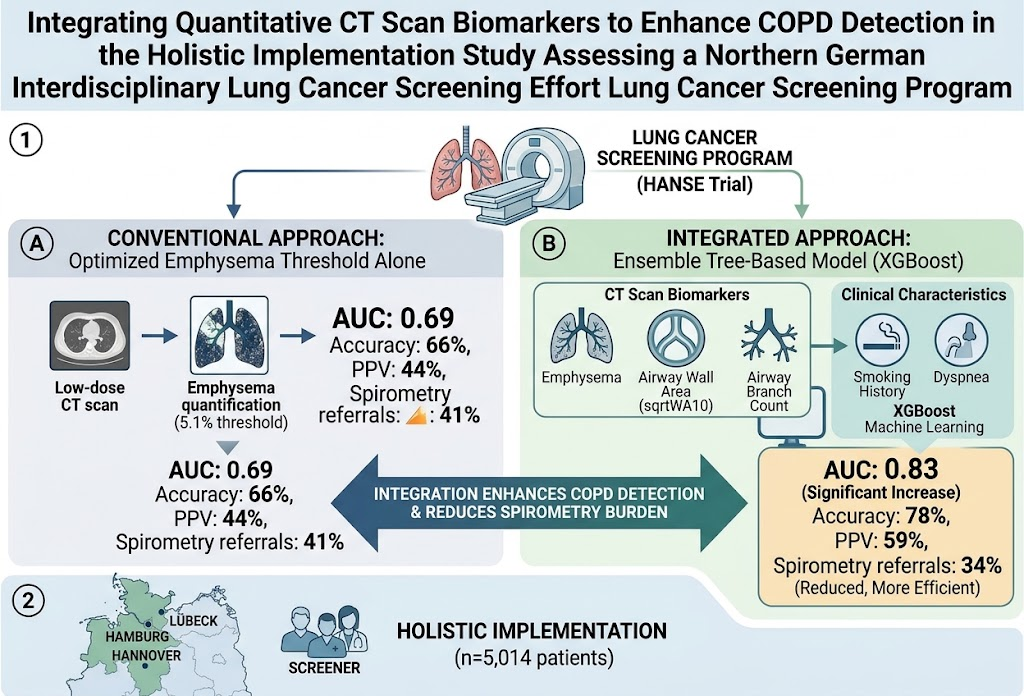
The Holistic Implementation Study Assessing a Northern German Interdisciplinary Lung Cancer Screening Effort enrolled more than 5,000 adults undergoing lung cancer screening, predominantly current or former smokers with ≥10 pack-years history. Spirometry was performed to identify previously undiagnosed COPD defined by airflow obstruction (FEV1/FVC < 0.70), with a sensitivity analysis applying lower limit of normal criteria. Low-dose chest CT scans were analyzed using artificial intelligence-based software to quantify emphysema extent, standardized airway wall thickness (square root wall area of airways with a theoretical internal perimeter of 10 mm), and airway branch count. An ensemble tree-based machine learning model—Extreme Gradient Boosting—integrated quantitative imaging biomarkers and clinical features such as smoking history and dyspnea to predict COPD presence. Diagnostic performance metrics were reported including area under the receiver operating characteristic curve (AUC), accuracy, positive predictive value (PPV), and the proportion of patients referred for confirmatory spirometry.
Key Findings
Among the 5,014 screening participants with spirometry data, 1,115 (22.2%) were found to have previously undiagnosed COPD. When analyzed without incorporating airway biomarkers, emphysema extent alone at an optimized threshold of 5.1% demonstrated modest discrimination of COPD (AUC 0.69; 95% CI, 0.67-0.72), correctly classifying 66% of patients, with a PPV of 44%. Notably, 41% of patients exceeded this threshold, triggering recommendation for confirmatory spirometry—representing a substantial referral burden.
The integrated machine learning model combining emphysema, airway wall thickness, airway branch count, and clinical characteristics significantly elevated diagnostic performance. It achieved an AUC of 0.83 (95% CI, 0.80-0.86), accuracy of 78%, and a PPV of 59%, while lowering referrals for confirmatory spirometry to 34% of patients. This approach thus improves detection efficiency by prioritizing higher-likelihood cases for spirometry confirmation.
In sensitivity analyses using the lower limit of normal spirometry criteria to define COPD, the integrated model’s performance further improved, with an AUC of 0.86 (95% CI, 0.83-0.89), accuracy of 84%, and PPV of 53%, with only 22% of patients referred for confirmatory testing. These findings underscore the robustness of quantitative CT airway biomarkers combined with clinical data in enhancing early COPD identification.
Expert Commentary
This study addresses a critical gap in COPD detection during lung cancer screening programs by leveraging advanced imaging biomarkers and machine learning. The integration of emphysema quantification with airway wall thickness and branching metrics reflects a more comprehensive assessment of COPD-related lung structural changes beyond emphysema alone. The observed improvement in positive predictive value and reduction in unnecessary spirometry referrals highlight the practical clinical relevance of this approach.
However, some limitations require consideration. The cohort predominantly comprises current and former smokers with significant tobacco exposure, potentially limiting generalizability to other populations. In addition, although CT-derived quantitative airway measures are promising, their availability and standardization outside research settings remain limited. Furthermore, while the machine learning model shows excellent discrimination, prospective validation and integration into clinical workflows are necessary to confirm real-world utility.
Current international COPD guidelines acknowledge the utility of CT imaging in disease phenotyping but have yet to formalize CT-based algorithms for screening or spirometry referral. Findings from this study may inform future recommendations by providing an evidence-based framework to leverage low-dose CT scans for opportunistic COPD identification and efficient resource allocation.
Conclusion
This study demonstrates that an integrated approach combining quantitative low-dose CT biomarkers of emphysema and airway morphology with clinical characteristics markedly enhances the detection of previously undiagnosed COPD in lung cancer screening cohorts. This strategy improves diagnostic accuracy, raises the positive predictive value for COPD, and decreases unnecessary referrals for confirmatory spirometry. Implementation of such models within lung cancer screening programs may enable targeted early COPD diagnosis, facilitating prompt clinical interventions and potentially reducing disease burden. Further studies are warranted to validate these findings across diverse populations and to evaluate long-term outcomes from integrated imaging-based COPD detection.
Funding and Clinical Trial Registration
The study was registered under ClinicalTrials.gov (NCT04913155). Detailed funding sources were not specified in the abstract. Full trial details can be accessed through ClinicalTrials.gov.
References
- Abdo M, Pott H, Reck M, et al. Integrating Quantitative CT Scan Biomarkers to Enhance COPD Detection in the Holistic Implementation Study Assessing a Northern German Interdisciplinary Lung Cancer Screening Effort Lung Cancer Screening Program. Chest. 2026 Aug 14. PMID: 42600771. https://pubmed.ncbi.nlm.nih.gov/42600771/
- Vestbo J, Hurd SS, Agusti AG, et al. Global strategy for the diagnosis, management, and prevention of chronic obstructive pulmonary disease: GOLD executive summary. Am J Respir Crit Care Med. 2013 Feb 15;187(4):347-65.
- GOLD Report 2023. Global Initiative for Chronic Obstructive Lung Disease. https://goldcopd.org/2023-gold-report-2/
- National Lung Screening Trial Research Team. Reduced lung-cancer mortality with low-dose computed tomographic screening. N Engl J Med. 2011 Aug 4;365(5):395-409.