Highlight
This article discusses the development and validation of a clinical likelihood score for identifying patients with pulmonary arterial hypertension (PAH) complicated by pulmonary veno-occlusive disease or pulmonary capillary hemangiomatosis (PVOD/PCH). Key highlights include:
- Patients with PVOD/PCH have worse outcomes and are underdiagnosed within Group 1 PAH.
- A novel scoring system incorporating clinical, functional, and imaging variables shows high accuracy (ROC AUC 0.97) in discriminating PVOD/PCH from other PAH forms.
- Early identification via this score can influence treatment decisions and expedite lung transplant referral.
Study Background
Group 1 pulmonary arterial hypertension (PAH) is a progressive disease marked by vascular remodeling leading to increased pulmonary vascular resistance and right heart failure. Within this group, a subset characterized by venous or capillary involvement, formerly classified as pulmonary veno-occlusive disease or pulmonary capillary hemangiomatosis (PVOD/PCH), exhibits overlapping clinical phenotypes but markedly different pathophysiology, prognosis, and treatment response.
PVOD/PCH patients often present with lower diffusing capacity of the lungs for carbon monoxide (DLCO), hypoxemia, and radiographic findings such as septal line thickening and lymphadenopathy. These patients are at higher risk of complications from conventional PAH vasodilator therapy, including pulmonary edema, and have substantially elevated mortality without timely lung transplantation.
Despite their clinical significance, PVOD/PCH cases remain underrecognized in practice due to the rarity of the condition, lack of specific noninvasive diagnostic tools, and the overlap with idiopathic PAH. The absence of large dedicated registries has hindered systematic study and diagnostic advancements.
Study Design
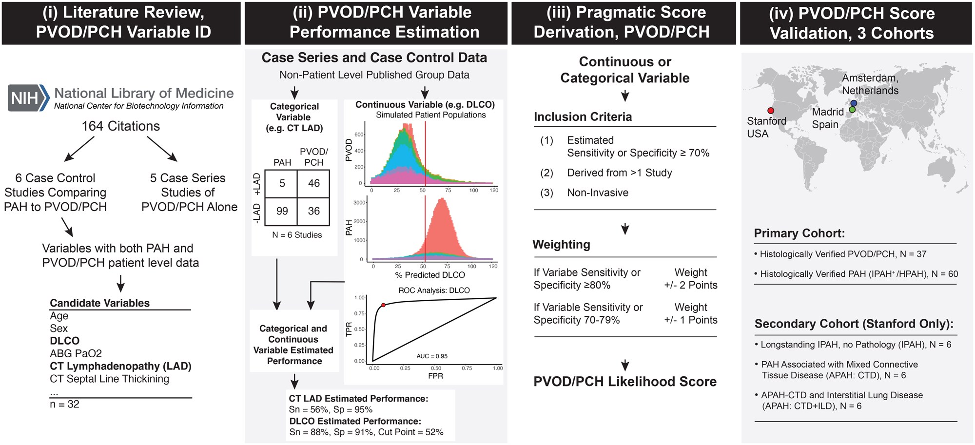
This investigation utilized a rigorous approach to develop a PVOD/PCH likelihood score by pooling data from published case-control and case-series studies focused on distinguishing PVOD/PCH from classical PAH. The authors extracted clinical and paraclinical variables showing discriminatory potential and performed sensitivity and specificity analyses, complemented by receiver operating characteristic (ROC) curve simulations to assess predictive accuracy.
The top-performing variables forming the composite score included DLCO, oxygen desaturation during the 6-minute walk test, arterial oxygen tension (PaO2), patient sex, smoking history, computed tomography (CT) findings of septal line thickening, and lymphadenopathy.
Validation cohorts comprised 97 lung transplant–eligible patients with histopathology-confirmed diagnosis—37 PVOD/PCH cases and 60 PAH controls—from centers in the United States, Spain, and the Netherlands, enabling assessment across geographic and demographic diversity.
Key Findings
The resulting PVOD/PCH likelihood score demonstrated exceptional diagnostic performance, yielding an area under the ROC curve of 0.97 (95% CI, 0.93–1.00), indicating near-perfect discrimination between PVOD/PCH and PAH. This accuracy was robust even when some data variables were missing, underscoring the score’s practical utility in real-world clinical scenarios.
Specifically, lower DLCO and PaO2 values, marked desaturation on the 6-minute walk test, and the presence of septal line thickening and lymphadenopathy on CT scans emerged as strong individual predictors. The inclusion of demographic factors such as male sex and smoking history improved the model’s discriminatory capacity.
This novel tool offers a pragmatic, evidence-based means to identify PVOD/PCH in incident PAH patients prior to therapy initiation—a critical step because standard PAH vasodilators may precipitate life-threatening pulmonary edema in this population.
Expert Commentary
This study addresses a significant clinical gap. Current guidelines acknowledge PVOD/PCH as a difficult diagnosis generally requiring histopathological confirmation post-lung transplantation or autopsy. The ability to noninvasively stratify patients at high likelihood of PVOD/PCH aids in personalized treatment planning and enhances transplant candidacy evaluation.
However, some limitations merit consideration. The reliance on pooled retrospective data and relatively small sample size, while understandable given disease rarity, could impact generalizability across more heterogeneous patient populations. Prospective validation in larger cohorts and integration with emerging biomarkers or genetic data could further refine predictive accuracy.
Clinicians should interpret the score alongside clinical context, recognizing that no test replaces comprehensive evaluation. Nonetheless, this tool forms a valuable adjunct to multidisciplinary PAH care teams.
Conclusion
The development of a validated PVOD/PCH likelihood score represents a meaningful advancement in PAH diagnostics. By integrating functional, radiographic, and demographic variables, the score enables early recognition of venous and capillary involvement, guiding safer therapy initiation and timely referral for lung transplantation.
Future work should focus on broader prospective validation, incorporation into clinical guidelines, and exploration of impact on patient-centered outcomes. Until then, adoption of this score can help bridge the current diagnostic gap in PVOD/PCH, ultimately improving prognosis in this high-risk group.
Funding and Clinical Trials
The original study was supported by institutional grants and collaborative funding from involved academic centers. No direct clinical trial registration was reported for this retrospective analysis.
References
- Andruska AM, Cruz-Utrilla A, Nossent EJ, et al. Identification of pulmonary arterial hypertension patients with venous or capillary involvement. Am J Respir Crit Care Med. 2026;212(9):2199-2210. PMID: 42275164.
- Galie N, Humbert M, Vachiery JL, et al. 2022 ESC/ERS Guidelines for the diagnosis and treatment of pulmonary hypertension. Eur Heart J. 2022;43(38):3618-3731.
- Boulate D, Humbert M. Pulmonary Veno-Occlusive Disease and Pulmonary Capillary Hemangiomatosis: An Update. Clinics in Chest Medicine. 2020;41(3):359-374.