Early Retinal Sensitivity Decline in Intermediate AMD: Insights from Lesion-Centered Functional Trajectories in the PINNACLE Study

Highlight

The prospective PINNACLE study reveals early and distinct patterns of retinal sensitivity loss in intermediate age-related macular degeneration (iAMD) localized to specific lesion types. Sensitivity decline precedes structural detection at ellipsoid zone/interdigitation zone loss sites, exhibits central retinal spatial vulnerability, and varies by lesion subtype. These findings support lesion-specific functional monitoring using microperimetry despite notable measurement variability.

Study Background

Age-related macular degeneration (AMD) is a leading cause of vision impairment globally, with intermediate AMD (iAMD) representing a stage where targeted interventions might slow progression. However, the relationship between structural retinal lesions and functional decline in iAMD remains incompletely understood. Specifically, early detection of localized retinal sensitivity loss relative to lesion development on optical coherence tomography (OCT) could improve outcome measures for emerging therapies and inform patient management. The PINNACLE study was designed to longitudinally assess lesion-centered microperimetry data, providing in-depth characterization of focal functional changes and measurement variability in iAMD.

Study Design

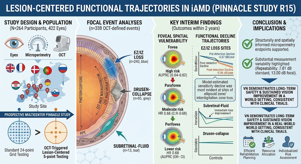
This report analyzes data from the prospective, longitudinal, multicenter PINNACLE study (NCT06682455), including 264 participants contributing 422 eyes in total. Microperimetry assessments used a mesopic 24-point standard grid for general functional mapping, augmented by a lesion-centered 5-point testing grid triggered by OCT-defined focal retinal events. The OCT events analyzed comprised ellipsoid zone/interdigitation zone (EZ/IZ) loss, drusen-collapse, and subretinal fluid occurrences. Repeatability of sensitivity measurements was evaluated using linear mixed-effects models. Spatiotemporal generalized additive mixed models estimated lesion-centered sensitivity trajectories by combining post-detection focal grid data with spatially interpolated pre-detection standard grid data. A secondary piecewise linear mixed-effects model quantified sensitivity changes before and after lesion detection.

Key Findings

Measurement Variability and Repeatability: The coefficient of repeatability for point-wise microperimetry was 7.61 dB (95% CI, 7.58-7.64) for the standard testing grid and increased to 13.00 dB (95% CI, 12.76-13.21) for the lesion-centered testing grid, indicating substantial measurement variability that must be accounted for in longitudinal functional assessment.

Event Types and Frequency: The study identified 338 OCT-defined lesion events in 131 eyes from 113 participants: 240 EZ/IZ loss, 85 drusen-collapse, and 13 subretinal fluid events.

Spatial Patterns of Sensitivity Loss: The risk of sensitivity loss ≥7 dB within two years was significantly lower at parafoveal (hazard ratio [HR] 0.66; 95% CI, 0.51-0.84) and perifoveal locations (HR 0.68; 95% CI, 0.53-0.87) compared to the fovea, demonstrating central retinal vulnerability in iAMD functional decline.

Lesion-Specific Sensitivity Trajectories:

  • EZ/IZ loss sites exhibited a measurable decline in sensitivity beginning before lesion detection at 0.57 dB/year (95% CI, 0.25-0.89) that accelerated post-detection, reaching 0.94 dB/year (95% CI, 0.55-1.34) in the later period.
  • Drusen-collapse sites did not demonstrate consistent longitudinal sensitivity decline, suggesting heterogeneous functional consequences.
  • Subretinal fluid events showed an immediate sensitivity loss of 2.18 dB (95% CI, 0.84-3.53), followed by early continued decline and some evidence of subsequent improvement, indicating complex dynamic functional changes.

Matched control retinal locations without lesions remained functionally stable over time.

Expert Commentary

The PINNACLE study provides critical insight into the nuanced, lesion-specific functional decline patterns in iAMD, underscoring the utility of spatially-informed microperimetry for early detection and monitoring. The observed pre-detection sensitivity loss at EZ/IZ loss sites suggests that functional changes can precede visible structural damage on OCT, potentially refining intervention timing. However, the considerable measurement variability highlights the ongoing challenges in employing microperimetry as a reliable clinical trial endpoint or biomarker in AMD.

These findings align with the emerging understanding that the foveal region is particularly susceptible to iAMD progression and functional deterioration, which might contribute to central vision loss impacting quality of life. The heterogeneous trajectories between lesion types support the need for tailored monitoring strategies. Future work incorporating larger cohorts, longer follow-up, and multimodal imaging could further elucidate the pathophysiological underpinnings of these functional patterns and optimize functional endpoints for therapeutic trials.

Conclusion

This study advances our knowledge of early retinal functional decline in iAMD by demonstrating lesion-centered and spatially-resolved sensitivity trajectories. The data emphasize central retinal vulnerability and distinct patterns of decline associated with EZ/IZ loss and other lesion types. Despite notable measurement variability, structurally informed microperimetry remains a promising modality to capture early retinal dysfunction, which could ultimately aid in earlier diagnosis, personalized monitoring, and evaluation of novel interventions in AMD.

Funding and ClinicalTrials.gov Information

The PINNACLE study is registered under NCT06682455. Funding sources were not specified in the abstract; further review of the full publication is recommended for detailed funding disclosures.

References

Futterknecht S, Riedl S, Mai J, et al. Lesion-Centered Functional Trajectories Reveal Early Retinal Sensitivity Decline in Intermediate AMD: PINNACLE Study Report 15. American Journal of Ophthalmology. 2026; in press. PMID: 42607745.

Additional relevant background literature includes:

  • Wong WL, Su X, Li X, et al. Global prevalence of age-related macular degeneration and disease burden projection for 2020 and 2040: a systematic review and meta-analysis. Lancet Glob Health. 2014;2(2):e106-16.
  • Schmidt-Erfurth U, Chong V, Loewenstein A, et al. Guidelines for the management of neovascular age-related macular degeneration by the EURETINA panel. Br J Ophthalmol. 2014;98(9):1144-67.
  • Pfau M, Sadda SR, Singh RP, Waldstein SM. Monitoring disease progression in intermediate AMD using multimodal imaging and microperimetry. Prog Retin Eye Res. 2021;83:100908.

Comments

No comments yet. Why don’t you start the discussion?

Leave a Reply