Highlight
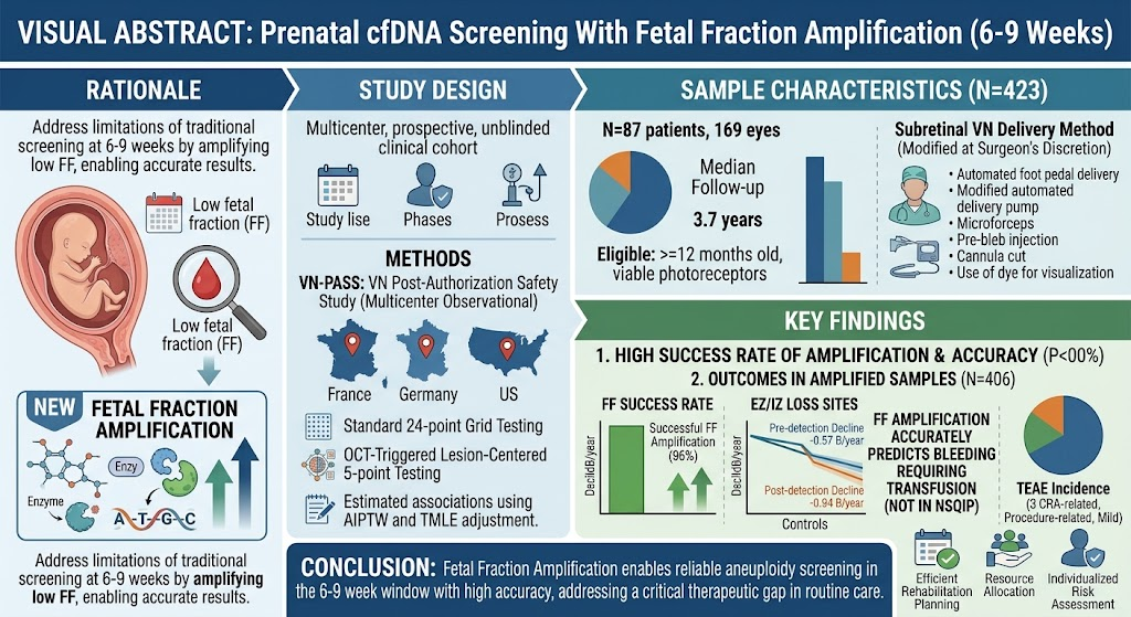
1. Fetal fraction amplification (FFA) enables prenatal cell-free DNA (cfDNA) screening as early as 7 3/7 weeks gestation, earlier than the traditional 10 weeks.
2. Using FFA, low fetal fraction rates dropped to 4.8% at 7 3/7 weeks, with screen failure rates as low as 0.3% after this point.
3. cfDNA screening between 8 0/7 and 9 6/7 weeks showed 100% concordance for aneuploidy detection and fetal sex determination compared to samples taken at 10 weeks or later.
4. Early screening with FFA may facilitate earlier prenatal risk assessment, benefiting clinical decision-making and patient counseling.
Study Background
Prenatal cell-free DNA screening is a widely adopted noninvasive test to screen for fetal chromosomal aneuploidies such as trisomy 21, 18, and 13. Standard cfDNA screening typically is performed at 10 weeks or later to ensure adequate fetal fraction—the proportion of cfDNA originating from the placenta compared to maternal DNA—which is crucial for reliable test accuracy. Low fetal fraction, often defined as less than 4%, can result in test failure or false-negative results. However, many pregnant individuals seek early risk assessment as soon as possible after confirming pregnancy to make timely decisions about testing and care.
Fetal fraction amplification is a novel molecular approach designed to increase the relative amount of fetal DNA in maternal blood samples, potentially enabling reliable cfDNA screening at earlier gestational ages when fetal fraction is naturally lower. Prior to this innovation, cfDNA screening before 10 weeks gestation faced challenging low fetal fraction rates, limiting its feasibility and clinical utility in very early pregnancy.
Study Design
This prospective observational study enrolled 562 pregnant individuals who underwent two blood draws: the first between 6 0/7 and 9 6/7 weeks of gestation and the second at 10 0/7 weeks or later. A cfDNA screening test incorporating fetal fraction amplification technology was applied to both samples.
The primary objective was to identify the earliest gestational age at which the proportion of samples with low fetal fraction (less than 4%) was reduced to 4.8% or less, a threshold based on historical data before fetal fraction amplification implementation.
The study assessed median fetal fraction, screen failure rates, positive and negative percent agreements for detection of common aneuploidies, and concordance of fetal sex determination between early and later gestational samples. Subgroup analysis examined the influence of maternal body mass index (BMI), given its known association with fetal fraction.
Key Findings and Results
A total of 470 participants completed both blood draws and were included in the final analysis. The median maternal age was 32 years, and median BMI was 25.7.
Fetal Fraction: Median fetal fraction for samples drawn between 6 0/7 and 9 6/7 weeks was 9.0%, considerably lower than the median 18.5% observed at 10 weeks or later but notably above the low-fetal fraction threshold in most cases. Among women with BMI ≥30 (n=125), fetal fractions were lower but similarly improved with later gestation and fetal fraction amplification (7.2% early vs. 14.7% later).
Gestational Age Threshold: Logistic regression identified 7 3/7 weeks as the earliest gestational age at which the proportion of low fetal fraction samples (defined as <4%) was 4.8% or less, matching historical baseline seen at 10 weeks pre-FFA.
Screen Failure Rate: At 6 0/7-9 6/7 weeks, the failure rate was 1.5%; this was reduced to 0.3% for samples obtained at 7 3/7 weeks or later. Considering the typical gestational age at initial prenatal care (8 weeks onwards), the screen failure rate between 8 0/7 and 9 6/7 weeks was very low at 0.4%, with both positive and negative percent agreements for detecting aneuploidies reaching 100% when compared to samples drawn at 10 weeks or later.
Aneuploidy Detection: For trisomy 21 and trisomy 7, two out of three positives identified at 10 weeks or later were concordantly positive in samples drawn between 8 0/7 and 9 6/7 weeks. No discrepancies were reported, indicating high reliability of early testing with fetal fraction amplification.
Fetal Sex Concordance: Concordance for fetal sex determination was perfect (100%) across 240 samples tested for both XX and XY calls, underscoring the technical validity of early cfDNA screening.
Expert Commentary
Fetal fraction amplification represents a significant advancement in cfDNA prenatal screening by addressing the perennial challenge of low fetal fraction in early pregnancy. The reduction in screen failure rates and maintenance of diagnostic accuracy at under 9 weeks gestation could transform clinical practice, allowing earlier risk stratification and potentially minimizing patient anxiety and the need for repeat testing.
However, the relatively limited sample size and performance data outside the 8-9 week window warrant cautious interpretation. Additional large-scale studies and real-world implementation data are needed to confirm these encouraging findings, verify cost-effectiveness, and evaluate impacts on downstream clinical management and outcomes.
Conclusion
This study provides compelling evidence that fetal fraction amplification enables reliable prenatal cfDNA screening as early as 7 3/7 weeks of gestation, with particularly robust performance from 8 weeks onward. The technology may significantly improve the feasibility of early prenatal genetic risk assessment, benefiting both patients and providers by delivering accurate, actionable information earlier in pregnancy.
As fetal fraction amplification becomes more widely studied and integrated, it holds the potential to shift prenatal screening paradigms toward earlier, noninvasive testing without compromising accuracy or increasing test failure rates.
Funding and Clinical Trials
Details on funding sources and trial registration were not reported in the abstract. Interested readers should consult the original publication in Obstetrics and Gynecology (2026;148(2):149-156) for more information.
References
Dugoff L, Mabey B, Bordwell J, Pierson S, Johansen Taber K, Muzzey D. Prenatal Cell-Free DNA Screening With Fetal Fraction Amplification at 6-9 Weeks of Gestation. Obstet Gynecol. 2026 May 21;148(2):149-156. doi:10.1097/AOG.0000000000004693. PMID: 42166774.