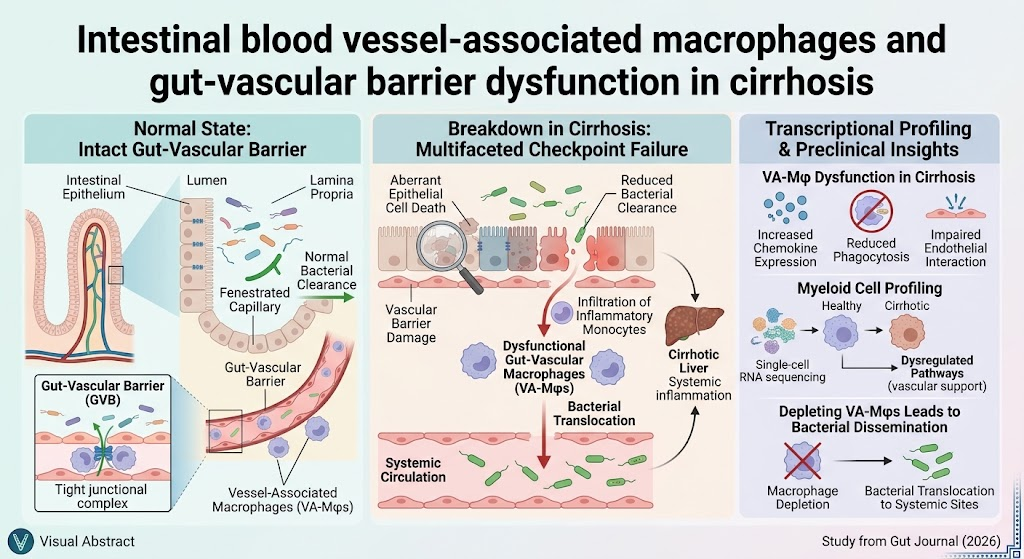
Highlight
- Bacterial translocation in cirrhosis involves multifactorial intestinal barrier disruption, including epithelial damage and vascular barrier dysfunction.
- Intestinal blood vessel-associated macrophages (vascular-lining macrophages) are critical for maintaining gut-vascular integrity and preventing systemic bacterial dissemination.
- Cirrhotic macrophages show impaired bacterial clearance, abnormal chemokine expression, and weakened vessel interactions, promoting infection risk.
- Translational data from patient duodenal biopsies confirm macrophage dysregulation observed in murine cirrhosis models, supporting clinical relevance.
Study Background
Cirrhosis, the advanced stage of chronic liver disease, is a major global health burden characterized by progressive liver fibrosis and portal hypertension. One of its critical complications is the increased risk of bacterial infections, often originating from the gut. Bacterial translocation—from the intestinal lumen through the mucosal barrier to mesenteric lymph nodes and systemic circulation—triggers systemic inflammation, precipitating hepatic decompensation, organ failure, and increased mortality. Despite recognition of the gut as the main bacterial source, the specific cellular and molecular mechanisms disrupting intestinal barrier function in cirrhosis remain incompletely understood.
The intestinal barrier involves the epithelial layer, the gut vascular barrier, and immune cells such as macrophages that patrol vascular and mucosal compartments. Recent research suggests that the interplay between these components is pivotal in preventing microbial breach. However, how vascular-associated macrophages contribute to barrier maintenance and how cirrhosis perturbs this system has remained elusive. Addressing this gap is critical for developing targeted strategies to prevent infections and their deleterious consequences in cirrhotic patients.
Study Design
Investigators developed a murine model of cirrhosis by administering carbon tetrachloride (CCl4) chronically over 20 weeks to induce liver fibrosis and cirrhosis features. This model allowed examination of the intestinal epithelial and vascular barriers. They isolated myeloid cells, particularly macrophages, from intestinal tissues in cirrhotic and control mice and conducted single-cell transcriptomic profiling to characterize functional alterations.
Complementing murine studies, the team analyzed duodenal biopsies from patients with compensated and decompensated cirrhosis to validate macrophage phenotypes and molecular changes. Functional assays evaluated macrophage bacterial clearance capacity and their spatial relationship with intestinal blood vessels, while experimental depletion of vascular-lining macrophages assessed their role in maintaining barrier integrity.
Key Findings
The study reveals that intestinal bacterial translocation in cirrhosis is not solely due to epithelial barrier damage but results from multifaceted breakdown involving epithelial cell death, vascular barrier compromise, and macrophage dysfunction.
Macrophage Dysfunction and Chemokine Dysregulation
Cirrhotic mice exhibited vascular-lining macrophages with elevated expression of monocyte-attracting chemokines, suggesting abnormal recruitment dynamics. Importantly, these macrophages showed reduced bacterial phagocytic capacity, impairing local bacterial clearance. Their altered interactions with blood vessel endothelium likely contribute to vascular barrier disruption.
Vascular Barrier Compromise
The study demonstrates that depletion of blood vessel-associated macrophages in otherwise healthy animals induced bacterial translocation, highlighting their essential role in maintaining gut-vascular barrier integrity independent of liver disease.
Translational Human Data
Single-cell RNA profiling of macrophages from duodenal biopsies of cirrhotic patients revealed transcriptional signatures echoing murine findings: downregulation of pathways that support vascular homeostasis, upregulation of chemokines, and signs of impaired functional capacity. These data confirm that gut-vascular macrophage dysfunction is a conserved feature in cirrhosis and likely contributes to increased systemic infection risk.
Expert Commentary
This study significantly advances our understanding of intestinal barrier pathophysiology in cirrhosis by focusing on the underappreciated gut-vascular macrophage compartment. Supporting prior evidence that gut microbial translocation drives cirrhosis complications, it uncovers a novel checkpoint involving vascular macrophages that actively maintain vascular integrity and clear bacteria.
The use of both an established preclinical model and patient-derived samples strengthens translational relevance. However, the complexity of cirrhosis pathogenesis necessitates further investigation into how systemic inflammation and portal hypertension interplay with macrophage dysfunction. These insights could lead to macrophage-targeted therapies or strategies enhancing gut-vascular barrier resilience to reduce infections without broad immunosuppression.
Current clinical approaches mostly focus on antimicrobial prophylaxis and managing portal hypertension; this work highlights the intestinal microenvironment as a promising therapeutic target. Future research should also examine whether reversing macrophage dysfunction or modulating chemokine profiles can restore barrier function and improve outcomes.
Conclusion
Bacterial translocation in cirrhosis arises from multifactorial intestinal barrier failures, including epithelial disruption, vascular barrier damage, and crucially, dysfunction of intestinal blood vessel-associated macrophages. These specialized macrophages are essential for maintaining the gut-vascular barrier, clearing bacteria, and preventing systemic dissemination.
This groundbreaking study using murine models and human biopsies provides compelling evidence that targeting macrophage pathways might offer new therapeutic avenues to prevent infections and their fatal sequelae in cirrhosis. Given the high morbidity and mortality associated with bacterial infections in this population, these findings hold significant clinical implications.
Continued exploration into gut-vascular immune homeostasis will be key to developing effective interventions to enhance intestinal barrier integrity and improve cirrhosis management strategies.
Funding and ClinicalTrials.gov
This research was supported by multiple academic and clinical institutions as cited in the original publication. No clinical trials are referenced in this preclinical and translational study.
References
Smets L, Viola MF, Boesch M, et al. Intestinal blood vessel-associated macrophages and gut-vascular barrier dysfunction in cirrhosis. Gut. 2026;75(10):1934-1949. doi:10.1136/gutjnl-2025-311900
Additional relevant literature:
– Sorribas M, Martin A, Staels B, et al. Intestinal barrier dysfunction in cirrhosis. Semin Liver Dis. 2021;41(1):43-56.
– Albillos A, Lario M, Álvarez-Mon M. Cirrhosis-associated immune dysfunction: distinctive features and clinical relevance. J Hepatol. 2014;61(6):1385-1396.