
Highlight
- Contact lens-associated fungal keratitis presents earlier and with fewer ocular comorbidities than non-contact lens cases.
- Similar fungal species profiles were seen across contact lens and non-contact lens groups, dominated by filamentous fungi.
- All contact lens-related cases resolved with medical therapy alone, whereas nearly a quarter of non-contact lens cases required surgical intervention.
- Final visual acuity outcomes were comparable regardless of contact lens use, emphasizing the importance of early diagnosis and treatment.
Study Background
Fungal keratitis is a serious corneal infection caused by fungi, leading to substantial ocular morbidity worldwide. It often results from ocular trauma, compromised ocular surface, or contact lens use. Contact lenses have been recognized as a risk factor for microbial keratitis, but their role in fungal keratitis and the clinical ramifications remain less well-defined. Understanding differential clinical features, microbiological profiles, and treatment outcomes in contact lens-associated and non-contact lens-associated fungal keratitis is essential for tailored management strategies and improving patient prognoses.
Study Design
This investigation was a retrospective, comparative clinical cohort study conducted at a tertiary referral center over eight years (2018–2025). Inclusion criteria encompassed patients with culture-proven fungal keratitis. Cases were stratified into two groups: contact lens-associated (CL) and non-contact lens-associated (non-CL). Data collection included demographics, ocular comorbidities, clinical presentation, microbiological identification of causative fungi, treatment regimens, and clinical outcomes such as resolution time, need for surgical intervention, and final visual acuity.
Key Findings
A total of 37 eyes were analyzed, with 16 cases in the CL group and 21 in the non-CL group. Significant findings included:
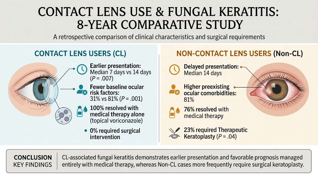
- Early Presentation: CL patients presented significantly earlier than non-CL patients (median 7 vs. 14 days; P = .007), likely reflecting heightened awareness among contact lens users or more acute symptom development.
- Underlying Risk Factors: Only 31% of CL cases had additional ocular risk factors compared to 81% in the non-CL group (P = .001), indicating that non-CL fungal keratitis frequently occurs in the presence of predisposing ocular surface disease or trauma.
- Microbiological Profile: Filamentous fungi predominated overall, including Fusarium species (16% overall; slightly more frequent in non-CL), Candida species (21% overall; slightly more in non-CL), and Aspergillus species (8%). The distribution did not significantly differ between groups, supporting that fungal etiology is similar regardless of contact lens status.
- Treatment Approaches: Topical voriconazole was the primary antifungal agent used in 78% of cases. All CL-associated fungal keratitis cases resolved with medical therapy alone, with a median healing time of 38 days (IQR 22-58). In contrast, 76% of non-CL cases resolved medically with a longer median healing time of 42 days (IQR 32-76), while 23% required therapeutic keratoplasty (P = .04), indicating a more complicated course.
- Visual Outcomes: Final best-corrected visual acuity was statistically comparable between both groups (logMAR 0.2 vs. 0.5; P = .56), suggesting that despite differing clinical courses, effective management can yield similar long-term vision preservation.
Expert Commentary
The analysis reveals distinct clinical trajectories influenced by contact lens use. Earlier diagnosis among CL patients likely facilitates prompt initiation of antifungal therapy, conferring improved outcomes and reducing the need for surgical intervention. The higher prevalence of ocular comorbidities in the non-CL group, including potentially chronic surface diseases or post-traumatic changes, may contribute to delayed presentations and poorer response to medical therapy alone.
While voriconazole remains a cornerstone in antifungal treatment, the study underscores the importance of individualized management depending on clinical context. Surgical intervention, though avoided in most CL cases, remains critical for complicated infections in the non-CL group. Clinical vigilance is necessary to identify patients at risk for therapeutic failure and to optimize timing of keratoplasty.
Limitations inherent to retrospective design include potential referral bias and variable treatment protocols. The relatively small sample size warrants cautious extrapolation. Future prospective studies with larger cohorts could elucidate mechanistic differences in fungal pathogenesis based on contact lens use and refine therapeutic algorithms further.
Conclusion
Contact lens-associated fungal keratitis differs clinically from non-contact lens cases by earlier patient presentation and fewer predisposing ocular conditions, both of which correlate with favorable resolution solely through medical therapy. Although fungal species distribution is similar regardless of contact lens use, non-contact lens-associated fungal keratitis carries a higher risk for complicated disease, often necessitating surgical intervention. Early recognition and appropriate antifungal treatment remain pivotal to optimizing visual outcomes in both groups.
Funding and ClinicalTrials.gov
The original study did not detail funding sources or registration with ClinicalTrials.gov.
References
1. Thomas PA, Kaliamurthy J. Mycotic keratitis: epidemiology, diagnosis and management. Clinical Microbiology and Infection. 2013;19(3):210–220. doi:10.1111/1469-0691.12134
2. Sharma N, Gopinathan U, Garg P, et al. Epidemiology and causative organisms of fungal keratitis in south India. Indian J Ophthalmol. 2002;50(2):77-82.
3. Whitcher JP, Srinivasan M. Corneal ulceration in the developing world—a silent epidemic. Br J Ophthalmol. 1997;81(8):622-623.
4. Prajna NV, Krishnan T, Rajaraman R, et al. The mycotic ulcer treatment trial: a randomized trial comparing natamycin vs voriconazole. JAMA Ophthalmol. 2013;131(4):422-429.