Highlight
This study demonstrates that airway mucus plugs detected on chest computed tomography (CT) are associated with increased exacerbation risk and lung function decline in individuals with chronic obstructive pulmonary disease (COPD), including those with preserved spirometry and those asymptomatic. Notably, a greater burden of mucus plugs correlates with higher risk of developing spirometry-defined COPD and more frequent moderate-to-severe exacerbations, suggesting mucus plugs as an early imaging biomarker for disease progression.
Study Background
Chronic obstructive pulmonary disease (COPD) represents a major global health burden characterized by persistent respiratory symptoms and airflow limitation. While mucus hypersecretion and plugging in airways are known to be hallmarks in advanced COPD and linked with poor outcomes, their role in earlier disease stages or in individuals without spirometric airflow obstruction has been less clear. Previous studies have largely focused on moderate-to-severe COPD, leaving a gap in understanding whether mucus plugs on chest CT can predict respiratory outcomes in people with preserved lung function or asymptomatic COPD. As COPD diagnosis often lags behind symptom onset and lung damage, identifying early imaging markers could improve prognostication and guide intervention to potentially slow disease progression.
Study Design
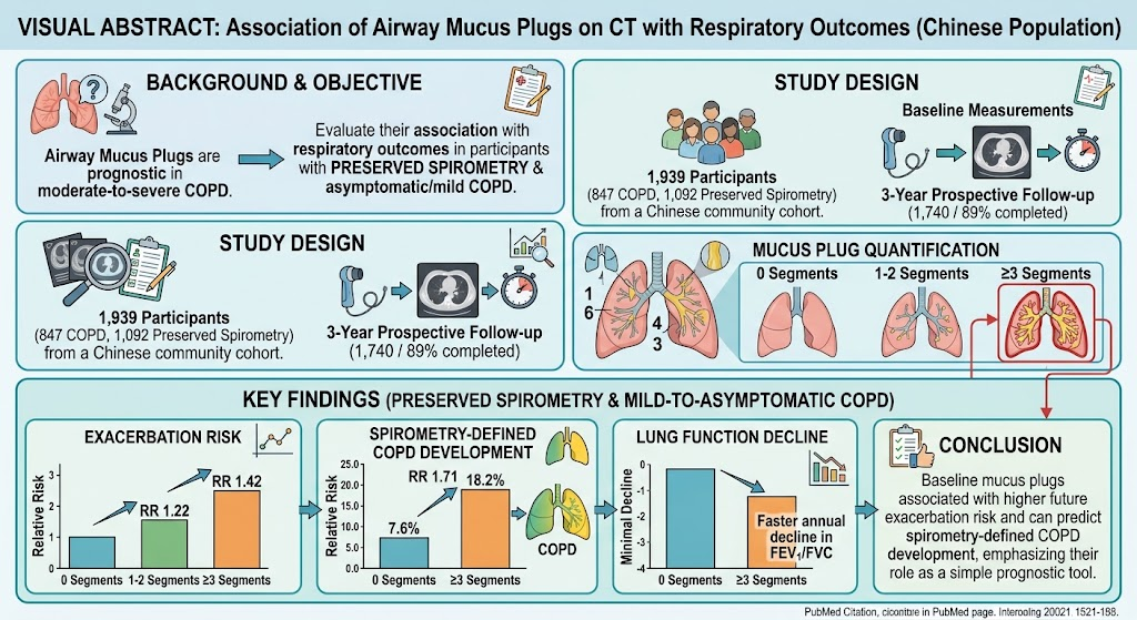
This investigation was a prospective, community-based cohort study conducted in China from July 2019 to September 2024 as part of the ECOPD study. The cohort included 1,939 participants subdivided into two groups: 847 with COPD (confirmed by spirometry) and 1,092 with preserved spirometry. Baseline assessments included standardized questionnaires to capture respiratory symptoms and exposures, spirometry tests, and chest CT scans. Mucus plugs were assessed on CT, and participants were categorized by the number of lung segments affected: 0, 1–2, or ≥3 segments. The primary respiratory outcomes evaluated over a 3-year follow-up were exacerbation frequency, annual lung function decline (particularly FEV1/FVC ratio), and progression to spirometry-defined COPD in the preserved spirometry group.
Key Findings
Among participants with preserved spirometry, having 1–2 mucus plug segments on baseline CT conferred a 22% increased risk for exacerbations (RR 1.22, 95% CI 1.01–1.48), while ≥3 plug segments further increased exacerbation risk to 42% (RR 1.42, 95% CI 1.11–1.81) compared to those without mucus plugs. The ≥3 segments subgroup also experienced a significantly higher rate of developing spirometry-defined COPD over 3 years (18.2% vs. 7.6%; RR 1.71, 95% CI 1.05–2.78) and a faster annual decline in the FEV1/FVC ratio, indicating accelerated airflow obstruction onset.
In participants with diagnosed COPD—particularly those with mild-to-moderate or asymptomatic disease—mucus plugs involving ≥3 lung segments were strongly associated with increased total exacerbations and moderate-to-severe exacerbations, further underscoring the relationship between mucus plugging and adverse clinical outcomes.
These findings collectively underscore that airway mucus plugs visible on CT scans serve as an independent prognostic indicator of worsened respiratory outcomes across the spectrum of COPD severity, including early or undiagnosed phases.
Expert Commentary
The study by Wu et al. contributes important evidence supporting the utility of CT imaging markers beyond traditional spirometry for COPD risk stratification and monitoring. The use of quantitative assessment of mucus plugging as a biomarker allows for identification of individuals at elevated risk for disease progression who might otherwise remain underrecognized due to preserved lung function or lack of symptoms. This aligns with emerging paradigms emphasizing early detection and targeted intervention before irreversible lung damage occurs.
However, the study is limited to a Chinese population, and external validation in diverse ethnic groups and geographic settings is necessary. Furthermore, integrating mucus plug assessment with other imaging and biomarker data will be critical to refine prognostic models and guide tailored therapies. Mechanistically, mucus plugs can exacerbate airflow obstruction by physically blocking small airways, promoting infection and inflammation, which may fuel disease progression.
Conclusion
Airway mucus plugs detected by chest CT provide clinically meaningful prognostic information for predicting respiratory outcomes, including exacerbations and development of spirometry-defined COPD in individuals with preserved lung function. This imaging biomarker could aid early identification of at-risk populations, allowing timely preventive strategies to mitigate disease advancement. Future research should explore the utility of routine mucus plug evaluation in COPD screening programs and its integration into clinical decision-making algorithms.
Funding and ClinicalTrials.gov
The study was supported by the ECOPD Investigators group and conducted under ethical oversight. Specific funding details were not disclosed in the abstract. The trial registration details were not provided but can be accessed in the original publication.
References
- Wu F, Wu F, Xia T, Zhou K, Deng Z, Wan Q, Tang G, et al. Association between Airway Mucus Plugs on Computed Tomography and Respiratory Outcomes in Participants with COPD and with Preserved Spirometry: Findings from a Chinese Population. Am J Respir Crit Care Med. 2026 Sep 25. PMID: 42789746.
- Global Initiative for Chronic Obstructive Lung Disease. Global Strategy for the Diagnosis, Management, and Prevention of COPD. 2024 Report. Available at: http://goldcopd.org
- Regan EA, et al. COPDGene Investigators. The genetic epidemiology of chronic obstructive pulmonary disease (COPD). Clin Chest Med. 2014 Sep;35(3):285-298.