Highlight
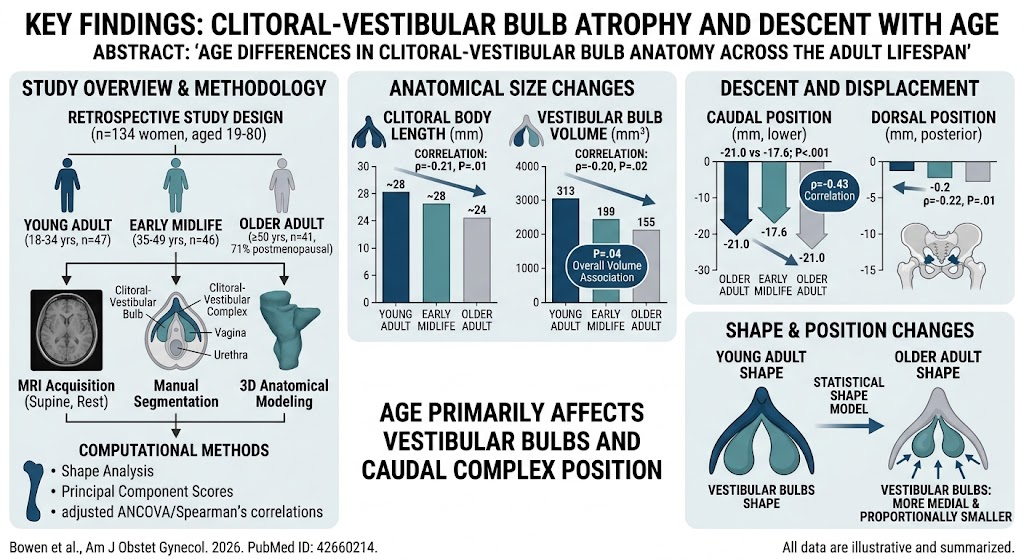
– Age-related atrophy and positional changes in the clitoral-vestibular bulb complex identified using 3D MRI models.
– Older women demonstrate smaller vestibular bulb volume and caudal displacement within the pelvis.
– Clitoral body length decreases moderately with age, whereas clitoral volume remains stable.
– Morphological alterations predominantly affect the vestibular bulbs, which become smaller and more medially positioned.
Study Background
The clitoral-vestibular bulb complex plays a critical role in female sexual function and contributes to pelvic structural integrity. Despite its clinical significance, normative anatomical data spanning the adult lifespan are sparse, particularly regarding age-related changes. This knowledge gap limits our understanding of how natural aging impacts female sexual health and pelvic symptoms such as dyspareunia and urinary complaints. With an increasing aging female population and the potential for age-related pelvic floor disorders, elucidating these anatomical changes is essential to inform clinical management and targeted therapies.
Study Design
This retrospective study analyzed pelvic magnetic resonance images (MRI) from 134 women aged 18 to 80 years without pelvic floor disorder symptoms or abnormalities on imaging. Participants were stratified into three age groups: Young Adult (18-34 years), Early Midlife (35-49 years), and Older Adult (≥50 years). Axial MRI images taken with subjects in the supine resting position were used for volumetric and positional assessment of the clitoral-vestibular bulb complex, vagina, and urethra. Manual segmentation facilitated construction of three-dimensional anatomical models. Morphometric parameters included clitoral body length, vestibular bulb volume, positional metrics (caudal, dorsal displacement), and shape analysis via principal component scores derived from a statistical shape model. Analyses accounted for confounders such as body mass index and parity using Bonferroni-corrected multivariate and univariate analyses of covariance. Spearman’s rank correlation assessed continuous age associations.
Key Findings
The median age of the cohort was 39 years; all women in the Young Adult and Early Midlife groups were premenopausal, whereas 71% of the Older Adult group were postmenopausal. Notable findings included:
- Older Adult women exhibited significantly shorter clitoral body length than Early Midlife counterparts (24.1 mm vs. 27.9 mm; P=0.006).
- Vestibular bulb volume decreased with age, reaching statistical significance (P=0.04), while clitoral volume did not change (P=0.73).
- The clitoral-vestibular bulb complex was positioned more caudally in Older Adult women compared to both younger groups (approximately -21 mm vs. -17.6 to -17.9 mm; P<0.001), indicating descent within the pelvic anatomy.
- Correlations across all ages showed a moderate negative association of age with clitoral body length (ρ=-0.21), vestibular bulb volume (ρ=-0.20), and more pronounced caudal displacement (ρ=-0.43).
- Shape analysis revealed the vestibular bulbs became more medially positioned and proportionally smaller relative to the clitoris with advancing age (ρ=-0.28; P=0.001), suggesting regional atrophic remodeling.
These outcomes suggest that aging leads to atrophic changes primarily in the vestibular bulbs with relative preservation of clitoral volume but shortening of the clitoral body. The positional descent may have functional implications given anatomical proximity to the urethra and vagina.
Expert Commentary
This study robustly characterizes normative anatomical changes in the clitoral-vestibular bulb complex using advanced imaging and computational modeling, filling a critical knowledge gap. The findings underscore that aging affects the vestibular bulbs more than the clitoris, potentially contributing to alterations in sexual response and pelvic floor dynamics. Given that a majority of Older Adult women in the cohort were postmenopausal, hormonal changes may mediate these effects, warranting further research.
Limitations include the retrospective design and imaging during rest without functional assessment. External factors such as hormone replacement therapy, sexual activity, and comorbidities were not extensively analyzed but could influence morphology. Future prospective studies incorporating sexual function outcomes and biomechanical analyses would better elucidate clinical relevance.
Conclusion
In conclusion, this study demonstrates that aging in women is associated with measurable anatomical changes in the clitoral-vestibular bulb complex, characterized by reduced vestibular bulb volume, shortened clitoral body length, and caudal pelvic descent. These changes may underlie some aspects of pelvic symptoms and sexual dysfunction seen in older women. Enhanced understanding of these age-related alterations may inform clinical evaluation and guide development of targeted therapies to improve female pelvic health across the lifespan.
Funding and ClinicalTrials.gov
Details regarding funding sources were not disclosed in the abstract. No clinical trial registration information was provided.
References
1. Bowen ST, Moalli PA, Dutta A, et al. Age Differences in Clitoral-Vestibular Bulb Anatomy Across the Adult Lifespan. American Journal of Obstetrics and Gynecology. 2026 Aug 27. PMID: 42660214.
2. Bachmann GA, Leiblum SR. The physiology of female sexual function. Archives of Sexual Behavior. 2004;33(2):179-186.
3. Palacios S, Lopez A, Aguirre V. Hormonal changes in the menopause transition and pelvic floor disorders: A review. Climacteric. 2020;23(2):179-186.
4. Rivas-Vazquez FJG, Edelman ER, Abramowitch SD. Advances in imaging female pelvic anatomy: implications for dysfunction diagnosis. Radiology Clinics. 2024;62(1):45-58.