Highlights
In this prospective cohort of patients with lacunar or mild nonlacunar stroke, large-artery stenosis of at least 50% was associated with lower odds of lacunar stroke and showed no meaningful association with MRI markers of cerebral small-vessel disease.
By contrast, basilar artery dolichoectasia and wider intracranial arteries were strongly associated with lacunar stroke phenotype, greater total cSVD burden, incident infarcts during follow-up, and progression of white matter hyperintensities over 1 year.
The findings support a predominantly nonatheromatous mechanism for lacunar stroke and cSVD, consistent with intrinsic microvascular injury rather than upstream embolic or flow-limiting large-artery disease.
Clinically, the study argues for mechanism-specific phenotyping in mild stroke and cautions against assuming that coexisting cranial arterial stenosis is the driver of lacunar infarction.
Background and Clinical Context
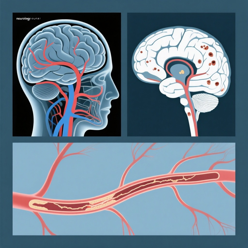
Cerebral small-vessel disease is a major cause of lacunar ischemic stroke, vascular cognitive impairment, gait dysfunction, mood symptoms, and intracerebral hemorrhage. On MRI, it commonly manifests as white matter hyperintensities, lacunes, enlarged perivascular spaces, microbleeds, and brain atrophy. Yet an important mechanistic question remains unsettled: to what extent are lacunar stroke and cSVD caused by intrinsic small-vessel pathology versus embolic or hemodynamic effects from large-artery disease?
This distinction matters clinically. If lacunar stroke often reflects parent or proximal arterial atherosclerosis, then vascular imaging findings such as intracranial or cervical stenosis would have direct etiologic and therapeutic implications. If, instead, cSVD primarily reflects diffuse arteriolar injury, endothelial dysfunction, vessel wall remodeling, and impaired autoregulation, then treatment priorities may shift toward blood pressure control, microvascular risk modification, and long-term brain health surveillance rather than stenosis-focused interventions.
Dolichoectasia adds another layer to this debate. The term refers to elongation, widening, and tortuosity of arteries, most classically the basilar artery. It has often been considered a marker of abnormal arterial wall structure and remodeling rather than classic atherosclerotic plaque burden. Whether this macrovessel phenotype signals parallel disease in the cerebral microcirculation has been uncertain. The current study directly addresses that question in a well-phenotyped prospective mild stroke cohort with baseline and follow-up MRI.
Study Structure and Methods
Study design
Han and colleagues conducted a prospective cohort study of patients with either lacunar stroke or mild nonlacunar ischemic stroke. Participants underwent detailed baseline phenotyping, including demographics, vascular risk factors, stroke subtype assessment, cognition and function measures, and MRI evaluation. Follow-up at 1 year included repeat imaging and clinical assessment, allowing the investigators to examine both cross-sectional and longitudinal associations.
Population
The cohort included 229 patients with a mean age of 65.9 years; 57.2% had lacunar stroke. This is an important design strength because the comparison group was not healthy controls but patients with another ischemic stroke phenotype, which helps address whether large-artery abnormalities are specifically enriched in lacunar disease.
Exposure definitions
Large-artery stenosis was defined as at least 50% stenosis in intracranial or cervical arteries. Basilar artery dolichoectasia was assessed using standard morphometric features, including basilar artery diameter, bifurcation height, and lateral displacement. The investigators also measured intracranial carotid and middle cerebral artery diameters, enabling analysis of a broader arterial-widening phenotype rather than a basilar artery-only effect.
Outcomes
The main outcomes included stroke subtype association, MRI markers of cSVD, total cSVD burden, incident infarcts over 1 year, and progression of white matter hyperintensities. The analysis also considered embolic sources and adjusted for age, sex, and vascular risk factors using multivariable logistic, linear, and proportional odds models.
Systematic review component
Beyond the cohort analysis, the authors performed a systematic literature review examining relationships between large-artery pathology and cSVD. This is useful because it places the cohort findings in the context of the broader evidence base rather than presenting them in isolation.
Key Results
Prevalence of arterial abnormalities
Among the 229 participants, large-artery stenosis was present in 20.5%, while basilar artery dolichoectasia was present in 15.7%. These are not trivial frequencies, underscoring that both narrowing and widening of cranial arteries commonly coexist in patients with mild stroke. The key question, however, is which vascular phenotype aligns with lacunar stroke biology.
Large-artery stenosis did not support a cSVD mechanism
After multivariable adjustment, large-artery stenosis was associated with lower odds of lacunar versus nonlacunar stroke, with an odds ratio of 0.49 (95% CI, 0.23-0.99). Put simply, patients with significant stenosis were less likely to have a lacunar stroke phenotype. The presence of any embolic source behaved similarly, again arguing against the notion that lacunar stroke in this cohort was primarily driven by upstream embolism or flow-limiting plaque.
Equally important, large-artery stenosis was not associated with cSVD markers or with incident infarcts during follow-up. This negative result has major interpretive value. It suggests that when significant intracranial or cervical stenosis is present in patients with mild stroke, it may often be an accompanying vascular comorbidity rather than the main driver of lacunar injury.
Basilar dolichoectasia showed a strong association with lacunar stroke
Basilar artery dolichoectasia showed the opposite pattern. It was strongly associated with lacunar stroke, with an adjusted odds ratio of 4.67 (95% CI, 1.87-13.14). This is a large effect size and one of the most clinically striking findings of the study. Rather than plaque-related narrowing, arterial widening and elongation appeared to be the macrovessel phenotype most closely aligned with lacunar stroke.
Association with overall cSVD burden
Basilar artery dolichoectasia was also associated with higher total cSVD scores, with an ordinal odds ratio of 2.57 (95% CI, 1.28-5.25). This suggests that the relationship is not limited to the index stroke subtype but extends to the broader brain MRI signature of chronic small-vessel injury. The finding is biologically coherent if dolichoectasia reflects diffuse abnormalities in vessel wall integrity or extracellular matrix remodeling that affect both large and small intracranial vessels.
Longitudinal imaging outcomes
One of the strengths of the paper is its longitudinal design. Basilar dolichoectasia was associated with incident infarcts over 1 year, with an odds ratio of 2.29 (95% CI, 1.01-5.14). Notably, 75% of these incident infarcts were subcortical, which again reinforces the link to small-vessel territory injury.
Basilar dolichoectasia was also associated with greater progression of white matter hyperintensities over 1 year, with a beta coefficient of 0.15 (95% CI, 0.01-0.29) per log10-transformed volume. Although the absolute numerical increment appears modest, white matter hyperintensity progression over a relatively short interval of 1 year is clinically meaningful and supports an active, ongoing disease process rather than a static anatomical variant.
Wider intracranial arteries behaved similarly
The pattern was not limited to the basilar artery. Similar associations were seen with larger intracranial carotid and middle cerebral artery diameters. This broadens the implication from a posterior circulation curiosity to a more generalized intracranial arterial-widening phenotype associated with cSVD.
Systematic review findings
The systematic review supported the cohort results, lending external consistency to the conclusion that arterial widening and dolichoectasia are more closely related to cSVD than atherosclerotic stenosis. In mechanistic research, convergence across study designs is particularly valuable because it reduces the likelihood that a single-cohort result is due to chance or local practice patterns.
How Should Clinicians Interpret These Findings?
The study challenges a common reflex in stroke medicine: attributing a small deep infarct to upstream large-artery disease simply because stenosis is visible on angiography. In many patients, especially older adults with vascular risk factors, stenosis and cSVD can coexist. This work suggests that coexistence should not be confused with causation.
For the practicing clinician, the message is nuanced but important. When a patient presents with a lacunar syndrome or subcortical infarct pattern, the presence of significant extracranial or intracranial stenosis should still be documented and managed according to established secondary prevention principles. However, it should not automatically displace intrinsic small-vessel disease as the leading etiologic explanation unless the infarct pattern, vessel territory, plaque morphology, or perfusion context clearly supports that mechanism.
By contrast, identification of basilar dolichoectasia or generalized intracranial arterial widening may carry greater relevance than previously appreciated. These features may serve as imaging markers of a diffuse arteriopathy associated with progressive cSVD, recurrent subcortical infarction, and white matter injury. That does not yet translate into a specific targeted therapy, but it may improve risk stratification and motivate closer longitudinal monitoring.
Biological Plausibility and Mechanistic Insight
The authors interpret their findings in favor of a nonatheromatous intrinsic microvascular pathology, particularly segmental arteriolar disorganization. This concept is plausible. cSVD is increasingly understood as a disorder of the small perforating arteries, arterioles, capillaries, and venules, involving endothelial dysfunction, blood-brain barrier leakage, smooth muscle cell loss, vessel stiffening, impaired vasoreactivity, and perivascular drainage failure.
Dolichoectasia may represent the large-artery counterpart of this abnormal vessel-wall biology. Rather than stenosis caused by focal plaque accumulation, widening and tortuosity may reflect structural weakness, matrix remodeling, elastin disruption, collagen abnormalities, or chronic pulsatile stress. If similar remodeling tendencies occur in the deep perforators, the result could be lipohyalinosis-like injury, impaired autoregulation, and vulnerability to lacunar infarction and white matter damage.
This mechanistic framework also helps explain why incident lesions in the study were predominantly subcortical. A diffuse arteriopathy would be expected to preferentially affect perforator-supplied regions rather than generate cortical territorial infarcts typical of large-artery embolism.
Strengths of the Study
Several features make this study particularly informative. First, it was prospective, with baseline and 1-year follow-up MRI, allowing evaluation of lesion accrual and white matter progression. Second, it directly compared lacunar and mild nonlacunar stroke phenotypes, which is more clinically relevant than comparing stroke patients with healthy controls. Third, it incorporated both stenotic and ectatic arterial phenotypes, avoiding an overly narrow atherosclerosis-centered framework. Fourth, the multivariable models adjusted for major confounders, including vascular risk factors. Finally, the embedded systematic review improves context and supports external validity.
Limitations and Cautions
As with any cohort study, causality cannot be proven. The association between dolichoectasia and cSVD may reflect shared upstream biology rather than a direct pathogenic sequence from large-artery widening to small-vessel injury. The sample size, although respectable for detailed MRI phenotyping, remains moderate, and confidence intervals for some estimates are relatively wide. Replication in larger multicenter cohorts would strengthen precision.
The study focused on patients with mild stroke, so generalizability to severe ischemic stroke, stroke-free populations, or primary prevention cohorts is uncertain. The abstract does not provide granular details about antithrombotic regimens, blood pressure trajectories, or differential management during follow-up, all of which could influence recurrent imaging findings. Also, arterial measurements may vary across imaging platforms and readers, so standardization will matter if these markers are to move toward broader clinical use.
Another practical limitation is that current stroke classification systems do not routinely give major weight to dolichoectasia or generalized intracranial arterial caliber when assigning mechanism. This paper suggests that such features may deserve more systematic consideration, but they are not yet integrated into guideline-based algorithms.
Relationship to Existing Literature
The findings align with the broader view that cSVD is primarily a diffuse arteriopathy rather than a downstream consequence of large-vessel atherosclerosis. The STRIVE recommendations established standardized neuroimaging markers for cSVD and helped frame it as a whole-brain vascular disorder rather than just a collection of incidental MRI lesions. Prior work from Wardlaw and colleagues has also emphasized endothelial and vessel-wall dysfunction, impaired reactivity, and tissue-level consequences across the brain.
At the same time, the paper does not argue that large-artery disease is irrelevant in stroke medicine. Rather, it refines the etiologic map: atherosclerotic stenosis matters greatly for many nonlacunar strokes, but it does not appear to explain most lacunar stroke or cSVD burden in this cohort. That distinction is clinically useful because overly broad attribution to “atherothrombotic disease” can blur treatment priorities and impede mechanistic trials.
Practice and Research Implications
For clinical practice, the immediate implication is improved diagnostic discipline. Radiologic evidence of significant stenosis should be interpreted in the context of infarct pattern and accompanying cSVD markers rather than treated as self-evident proof of mechanism. Conversely, dolichoectasia and enlarged intracranial arterial calibers may deserve explicit reporting when evaluating lacunar stroke and diffuse white matter injury.
For research, several next steps are apparent. Larger prospective cohorts should test whether arterial-widening phenotypes independently predict cognitive decline, gait impairment, recurrent stroke, or hemorrhage. Advanced vessel-wall imaging, hemodynamic studies, and blood or CSF biomarkers could help distinguish structural remodeling from inflammatory or degenerative pathways. Genetic and matrix biology studies may also be informative, especially in patients with marked dolichoectasia or disproportionate cSVD burden.
Most importantly, therapeutic development should become more mechanism-specific. If intrinsic microvascular pathology is the main driver, then interventions aimed at endothelial health, vascular stiffness, pulsatility, and blood-brain barrier integrity may be more relevant than escalating plaque-focused strategies in typical lacunar stroke populations.
Conclusion
This prospective study provides a clear and clinically meaningful message: cerebral small-vessel disease, including lacunar stroke, was not related to large-artery stenosis but was strongly associated with basilar dolichoectasia and wider intracranial arteries. The pattern supports a nonatheromatous intrinsic arteriopathy as the dominant mechanism of cSVD. For clinicians, the work sharpens etiologic reasoning in mild stroke. For researchers, it points toward a microvascular remodeling model that may better guide future diagnostics, risk stratification, and targeted therapy.
Funding and Trial Registration
The abstract provided does not report a ClinicalTrials.gov registration number. Specific funding details should be confirmed from the full Circulation publication.
References
1. Han F, Clancy U, Arteaga-Reyes C, Thrippleton MJ, Valdés Hernández MDC, Jaime Garcia D, Stringer MS, Backhouse E, Chappell FM, Cheng Y, Liu DX, Zhang J, Jochems ACC, Sakka E, Jardine C, Barclay G, McIntyre D, Hamilton I, Brown R, Zhu YC, Doubal FN, Wardlaw JM. Implications of Cranial Arterial Stenosis and Dolichoectasia for Cerebral Small-Vessel Disease Etiopathogenesis: Findings From a Prospective Mild Stroke Cohort. Circulation. 2026;153(23):1813-1826. PMID: 42090170.
2. Wardlaw JM, Smith EE, Biessels GJ, Cordonnier C, Fazekas F, Frayne R, Lindley RI, O’Brien JT, Barkhof F, Benavente OR, Black SE, Brayne C, Breteler M, Chabriat H, DeCarli C, de Leeuw FE, Doubal F, Duering M, Fox NC, Greenberg S, Hachinski V, Kilimann I, Mok V, Oostenbrugge R, Pantoni L, Speck O, Stephan BCM, Teipel S, Viswanathan A, Werring D, Chen C, Smith C, van Buchem M, Norrving B, Gorelick PB, Dichgans M. Neuroimaging standards for research into small vessel disease and its contribution to ageing and neurodegeneration. Lancet Neurol. 2013;12(8):822-838. PMID: 23867200.
3. Wardlaw JM, Smith C, Dichgans M. Mechanisms of sporadic cerebral small vessel disease: insights from neuroimaging. Lancet Neurol. 2013;12(5):483-497. PMID: 23602162.