Highlight
– Venous anatomy around the nipple-areolar complex (NAC) is characterized by a polygonal venous network critical for venous drainage.
– Venous injury quantified by vein branch loss (VBL) correlates with increased risk of significant NAC necrosis after nipple-sparing mastectomy (NSM).
– Higher mastectomy specimen weight, elevated BMI, and diabetes are additional risk factors.
– Near-infrared (NIR) imaging for venous visualization complements indocyanine green angiography (ICGA) in perioperative risk stratification and surgical planning.
Study Background
Nipple-sparing mastectomy (NSM) with immediate implant-based breast reconstruction (IBBR) is increasingly favored for its superior aesthetic and psychological outcomes compared with more radical mastectomy techniques. However, a substantial complication limiting its broader adoption remains the risk of nipple-areolar complex (NAC) necrosis. Preservation of NAC viability is paramount for optimal reconstructive success and patient satisfaction.
Historically, focus has been directed towards arterial insufficiency as the principal cause of NAC necrosis, with intraoperative perfusion assessment primarily relying on arterial imaging modalities such as indocyanine green angiography (ICGA). Nevertheless, venous drainage pathways are equally critical for tissue viability, and venous compromise may contribute to ischemia and necrosis but has been less thoroughly elucidated.
This study addresses an important gap by mapping the venous anatomy of the NAC, quantifying venous injury during NSM, and evaluating its clinical implications.
Study Design
The investigation comprised two complementary components: a cadaveric anatomical study and a prospective clinical cohort analysis.
The cadaveric study involved detailed dissection focusing on the superficial periareolar venous network to characterize the venous anatomy surrounding the NAC.
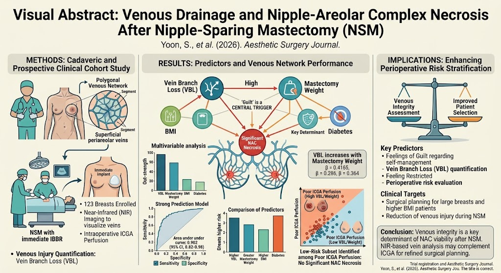
In the clinical cohort, 123 breasts undergoing NSM with immediate IBBR were prospectively evaluated. Near-infrared (NIR) imaging was employed intraoperatively to visualize cutaneous NAC veins. Venous injury was quantified objectively by calculating vein branch loss (VBL), defined as the extent of venous branches lost or compromised during mastectomy.
The primary clinical endpoint was the incidence of clinically significant NAC necrosis. Additionally, patient-related factors (body mass index [BMI], diabetes) and procedural variables (mastectomy specimen weight) were recorded. A prediction model was developed to integrate these factors and venous injury data, with its performance compared against ICGA findings in a subgroup of 32 breasts.
Key Findings
Venous Anatomy: Cadaveric dissection revealed a polygonal venous network immediately adjacent to the areolar margin, underscoring the complex and rich venous drainage system critical for NAC viability.
Clinical Outcomes: Significant NAC necrosis occurred in 19 out of 123 breasts (15.4%). On univariate analysis, higher VBL, increased mastectomy weight, elevated BMI, and presence of diabetes were all statistically associated with NAC necrosis (p < 0.05).
Multivariable modeling: Demonstrated that the risk associated with VBL was augmented in breasts with higher mastectomy weights, suggesting an interaction between venous injury severity and breast tissue volume removed.
Prediction Model Performance: The final multivariable risk prediction model yielded an area under the receiver operating characteristic curve (AUC) of 0.902 (95% confidence interval 0.82–0.98), indicating excellent discriminatory capability for clinically significant NAC necrosis.
Importantly, the model identified a low-risk subpopulation among breasts with poor ICGA perfusion where no significant NAC necrosis was observed, highlighting the complementary value of venous assessment alongside arterial imaging.
Expert Commentary
This study advances understanding of NAC viability by shifting some focus from the well-studied arterial supply to the equally critical venous drainage system. The polygonal venous architecture described through cadaveric dissection provides an anatomical basis that explains how venous injury during mastectomy can compromise tissue survival.
The use of NIR imaging for real-time visualization and quantification of venous integrity represents an innovative intraoperative tool. Notably, these findings advocate for an integrated vascular assessment strategy combining both venous and arterial perfusion monitoring to better predict and prevent NAC necrosis.
While ICGA remains a gold standard for assessing arterial perfusion, it overlooks venous flow impairments that can lead to venous congestion and ischemic injury. This study’s data suggest that incorporating quantitative venous imaging may improve perioperative decision-making, potentially altering flap design, surgical dissection planes, or immediate postoperative management to mitigate necrosis risk.
Limitations include the study’s observational design and a moderately sized clinical cohort. Larger multi-center investigations are warranted to validate these findings and to assess the generalizability across diversity in surgical technique and patient populations.
Conclusion
Venous integrity, as measured by vein branch loss through near-infrared imaging, is a critical determinant of nipple-areolar complex viability after nipple-sparing mastectomy with immediate reconstruction. This study’s prediction model demonstrates high accuracy in identifying patients at risk for NAC necrosis and exposes a subset of patients with poor arterial perfusion but preserved venous drainage who remain at low risk.
Integrating quantitative venous assessment with existing arterial imaging modalities may enhance perioperative risk stratification, surgical planning, and ultimately improve functional and aesthetic outcomes for patients undergoing NSM.
Funding and Registration
No specific funding information was provided in the original article abstract. The study was registered and reported under PMID: 42336360.
References
1. Yoon S, Park SO, Imanishi N, Zhu Y, Chang H. Venous Drainage and Nipple-Areolar Complex Necrosis After Nipple-Sparing Mastectomy: A Cadaveric and Prospective Clinical Cohort Study. Aesthet Surg J. 2026 Sep 16;46(10):1127-1136. PMID: 42336360.
2. Coopey SB, Tang R, Lei L et al. Nipple-sparing mastectomy and reconstruction: complications and outcomes. Ann Surg Oncol. 2013;20(10):3229-3235.
3. Moyer HR, Ho AL, Stempel M et al. Identification of risk factors for nipple-areola complex necrosis after nipple-sparing mastectomy. Ann Surg Oncol. 2012;19(6):1913-1918.
4. Newman MI, Froom J, Goldberg NH. Indocyanine green fluorescence mapping for breast reconstruction and surgical planning. Plast Reconstr Surg. 2009;124(3):726-733.