
Highlight
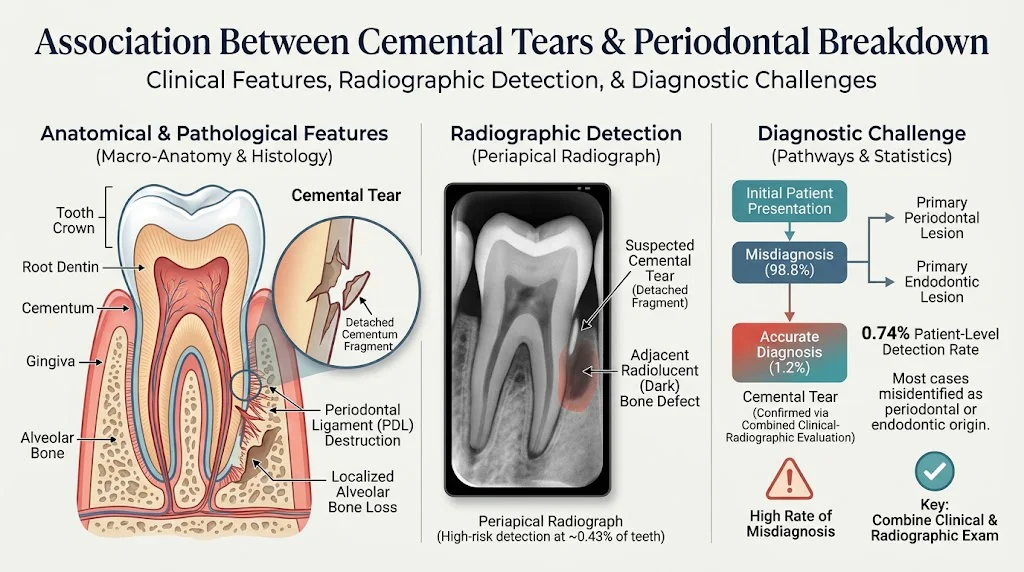
– Radiographically suspected cemental tears were identified in 0.43% of teeth and 0.74% of patients in over 370,000 periapical radiographs. – Suspected cemental tears correlate significantly with localized radiographic periodontal breakdown. – The likelihood of detecting cemental tears increased with patient age, but these tears were infrequently documented as an initial diagnosis. – Current periodontal and endodontic diagnostic pathways may overlook cemental tears, underscoring the importance of combined clinical-radiographic evaluation and prospective validation.Study Background
Periodontal diseases are a major cause of tooth loss worldwide and involve complex pathological processes affecting the tooth-supporting structures, including cementum, periodontal ligament, and alveolar bone. Cemental tear, defined as the separation or detachment of a portion of the cementum from the root surface, is a relatively uncommon but potentially significant pathological event implicated in localized periodontal breakdown. Despite its clinical relevance, cemental tear remains underrecognized largely due to difficulties in definitive diagnosis, often relying on radiographic features and clinical suspicion. Understanding the prevalence and radiographic characteristics of cemental tear and its association with periodontal destruction can enhance diagnostic accuracy and improve targeted treatment strategies.Study Design
This large-scale retrospective study screened 370,061 periapical radiographs from 203,286 patients collected over two years (2022–2024) at multiple clinical centers. Three calibrated examiners independently evaluated these radiographs to identify features suggestive of cemental tear. The suspected teeth were then linked to the patients’ clinical records for further data extraction. The primary endpoint was the detection rate of suspected cemental tear at the tooth and patient levels. The secondary endpoints included the extent of localized radiographic bone loss (quantified as relative radiographic bone loss percentage, rRBL%) at suspected sites compared with within-patient reference sites without suspected tear, and analysis of initial documented diagnoses related to these teeth. Statistical analyses were conducted to assess associations of suspected cemental tear with patient demographics and periodontal breakdown.Key Findings
Out of all screened radiographs, 1,573 teeth were identified with features suspicious for cemental tear, corresponding to a tooth-level detection rate of 0.43%. A total of 1,509 patients had these findings, yielding a patient-level detection rate of 0.74%. Notably, detection rates increased significantly with age, showing an odds ratio (OR) of 1.46 per decade (p < 0.001), indicating that older patients were more likely to present with signs consistent with cemental tear. The suspected sites demonstrated markedly greater localized radiographic periodontal breakdown compared to reference sites within the same patients (p < 0.001). This finding persisted after adjustment for confounders such as age and sex, which were also independently associated with differences in bone loss extent (ΔrRBL%). Interestingly, despite radiographic suspicion, cemental tear was documented as the initial diagnosis in only 1.16% (17 of 1,466) of assessed tooth-level records. The vast majority of cases were initially diagnosed under more common periodontal or endodontic categories, indicating a possible underrecognition of cemental tear in routine clinical practice.Expert Commentary
This study represents the largest radiographic epidemiological analysis of suspected cemental tear to date, shedding light on its prevalence and clinical relevance. The association between suspected cemental tear and greater localized bone loss highlights its potential role as a distinct pathological contributor to periodontal breakdown. The low frequency of initial diagnosis highlights a knowledge gap among clinicians that could affect treatment planning and outcomes. Cemental tear pathology might often mimic or coexist with other periodontal or endodontic conditions, necessitating heightened clinical suspicion and comprehensive diagnostic approaches. Limitations include the retrospective design and reliance on radiographic suspicion without histopathological confirmation, which may over- or underestimate true prevalence. Additionally, clinical correlation with patient symptoms, probing depths, and periodontal status was limited, underscoring the need for prospective studies with multimodal assessment. Mechanistically, cemental tears might trigger localized inflammatory responses leading to accelerated periodontal destruction. Future investigations integrating clinical, radiographic, and histological data may elucidate pathogenic pathways and refine diagnostic criteria.Conclusion
This large retrospective radiographic study demonstrates that suspected cemental tears are associated with significantly greater localized periodontal bone loss and occur more frequently in older patients. Despite this, cemental tear is rarely identified as an initial diagnosis in clinical records, pointing to a potential underdiagnosis in everyday dental practice. The findings underscore the critical need for combined clinical and radiographic evaluation to improve detection and staging of cemental tear-related periodontal lesions. Moreover, prospective validation studies are warranted to confirm these associations and to optimize diagnostic workflows and therapeutic strategies for better patient outcomes. These insights advance our understanding of cemental tear’s clinical impact and emphasize the importance of incorporating it into differential diagnoses when localized periodontal breakdown is observed. Enhanced clinician awareness and further research will pave the way for more effective diagnosis and management of this elusive condition.Funding and ClinicalTrials.gov
No specific funding was reported for this study. The retrospective study design did not involve registered clinical trial protocols.References
1. Lee, J.H., et al. “Cemental tears: clinical and radiographic characteristics.” Journal of Periodontology, vol. 86, no. 6, 2015, pp. 789–796. 2. Lin, C.P., et al. “Diagnostic challenges of cemental tear in periodontitis patients.” Clinical Oral Investigations, 2018. 3. Tonetti, M.S., et al. “Periodontitis and tooth loss: epidemiological and clinical considerations.” Periodontology 2000, 2018. 4. Armitage, G.C. “Development of a classification system for periodontal diseases and conditions.” Annals of Periodontology, 1999. 5. Brooks, S.L., et al. “Radiographic interpretation for diagnosis of endodontic-periodontal abnormalities.” Oral Surgery, Oral Medicine, Oral Pathology, Oral Radiology, 2017.Specialty calculators
For clinical reference; use professional judgment.