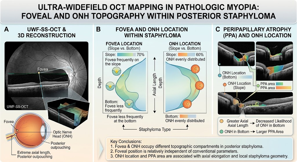
Ultra-widefield swept-source OCT (UWF-SS-OCT) enables simultaneous 3D assessment of the fovea, optic nerve head (ONH), and posterior staphyloma contour in pathologic myopia.
The fovea and ONH often occupy different topographic regions within the posterior staphyloma, indicating heterogeneous posterior pole remodeling.
Optic nerve head location correlates inversely with axial length; longer eyes show less ONH presence at the staphyloma bottom.
Peripapillary atrophy area associates with both axial length and ONH location, underscoring distinct mechanisms underlying macular and peripapillary changes in pathologic myopia.