

Introduction
Age-related macular degeneration (AMD) is a leading cause of vision loss primarily affecting the central retina, or macula, especially in older adults. A hallmark of advanced AMD is the development of atrophic lesions where the retinal pigment epithelium (RPE) and outer retina progressively degenerate. Understanding the precise growth dynamics of these atrophic areas is critical for prognostication and for guiding therapeutic approaches. Previous studies have recognized different precursor lesion types that precede atrophy, including drusenoid deposits, subretinal drusenoid deposits (SDD), and acquired vitelliform lesions (AVL). However, it remains unclear whether these distinct precursors give rise to atrophic lesions with different growth rates and temporal patterns.Study Objectives
This retrospective longitudinal cohort study aimed to evaluate if atrophic lesions emerging from various retinal precursor types demonstrate distinct temporal kinetics in their growth after the onset of complete retinal pigment epithelium and outer retinal atrophy (cRORA) in patients with intermediate AMD (iAMD). Clarifying these patterns may provide new insights into disease progression pathways and aid in patient stratification for clinical trials and management.Methods
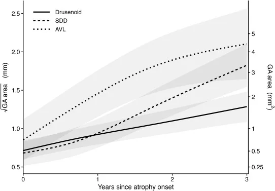
The study reviewed 109 eyes from 88 patients with intermediate AMD who progressed to cRORA. Using serial spectral-domain Optical Coherence Tomography (OCT) scans, researchers retrospectively identified the onset site of cRORA and traced back to characterize the precursor lesion present at that exact location. Precursors were classified into three types: drusenoid, subretinal drusenoid deposit (SDD), and acquired vitelliform lesion (AVL). Atrophic areas were automatically segmented on en face OCT projections and manually corrected for accuracy. Growth kinetics of the square-root-transformed total atrophic area were analyzed across the first three years following cRORA onset. A piecewise linear regression model with time knots at 1 and 2 years was used, adjusting for initial atrophic area, lesion eccentricity relative to the fovea, and number of foci at onset.Key Findings
Among the eyes studied, 66 progressed from drusenoid lesions, 31 from SDD, and 12 from AVL. Significant differences in growth rates of atrophy were observed based on precursor type: – Drusenoid group showed a steady growth rate of 0.197 mm/year. – SDD group demonstrated a faster growth rate of 0.383 mm/year. – AVL group exhibited the fastest growth at 0.429 mm/year. Both SDD and AVL groups expanded significantly faster than the drusenoid group (P<0.001), although the difference between SDD and AVL was not statistically significant. Temporal kinetic profiles diverged notably: – The drusenoid group exhibited relatively steady atrophic expansion across all three years. – The SDD group showed similar growth to the drusenoid in the first year but accelerated markedly during years two and three. – The AVL group grew fastest in the first year, with growth rate decelerating in subsequent years. These distinct growth trajectories remained robust after controlling for confounding factors such as initial lesion characteristics. Furthermore, the burden of parafoveal SDD outside the primary lesion site did not significantly affect growth rate once precursor lesion type was accounted for, highlighting the importance of the specific origin of the lesion.Clinical Implications
Identifying the nature of precursor lesions in AMD could serve as an important prognostic marker. Patients whose atrophy originates from drusenoid lesions may experience slower, more consistent progression, while those with SDD or AVL precursors may anticipate faster or temporally variable atrophic enlargement. Such stratification can inform patient counseling, monitoring frequency, and tailored interventions. In clinical trials testing novel treatments aimed at halting or slowing atrophy, incorporating precursor lesion types as stratification variables might improve the precision of outcome assessments and therapeutic responses. Moreover, recognizing distinct kinetics aids in understanding pathophysiological mechanisms governing each lesion subtype.Expanded Background and Context
AMD affects millions globally, with atrophic AMD comprising a significant and challenging subtype due to its relentless progression and lack of widely effective treatments. The drusenoid precursor is characterized by yellowish extracellular deposits beneath the RPE, historically associated with classic pathways of AMD progression. Subretinal drusenoid deposits (SDD), also called reticular pseudodrusen, accumulate above the RPE and have been increasingly recognized as a distinct and high-risk phenotype with strong links to geographic atrophy. Acquired vitelliform lesions (AVL) are characterized by yellowish subretinal material resembling that found in vitelliform dystrophies and may represent another atrophic progression pathway. Clarifying how these lesions influence subsequent atrophy growth kinetics is vital for a nuanced understanding of AMD heterogeneity, which impacts prognosis and therapeutic development.Future Directions and Recommendations
Further prospective studies with larger, ethnically diverse cohorts and advanced imaging biomarkers are necessary to validate and extend these findings. Integration of functional vision assessments and genetic profiling may also enhance prognostic accuracy. Interventions targeting specific pathways active in SDD or AVL-associated atrophy could be an avenue for personalized AMD treatment. Additionally, routine clinical imaging protocols should aim to identify precursor lesion types to better stratify patients.Conclusion
This study elegantly demonstrates that precursor lesion types in intermediate AMD significantly influence the rate and temporal pattern of atrophic lesion growth following cRORA onset. Drusenoid lesions precipitate more gradual and steady atrophy, while SDD lesions are associated with accelerated expansion later, and AVL lesions exhibit rapid early growth that subsequently slows. These findings hold promise for improving prognostic precision and designing targeted clinical trials in AMD management.Specialty calculators
For clinical reference; use professional judgment.