Proposed section structure
For this topic, a clinically and scientifically appropriate structure is: Highlights; Clinical background and unmet need; Study design and translational platform; Mechanistic findings; In vivo cardiac remodeling results; Human translational relevance; Clinical interpretation and limitations; Conclusion; Funding and trial registration; References. This structure matches the article’s blend of molecular immunology, post-myocardial infarction remodeling, and translational cardiovascular research.
Highlights
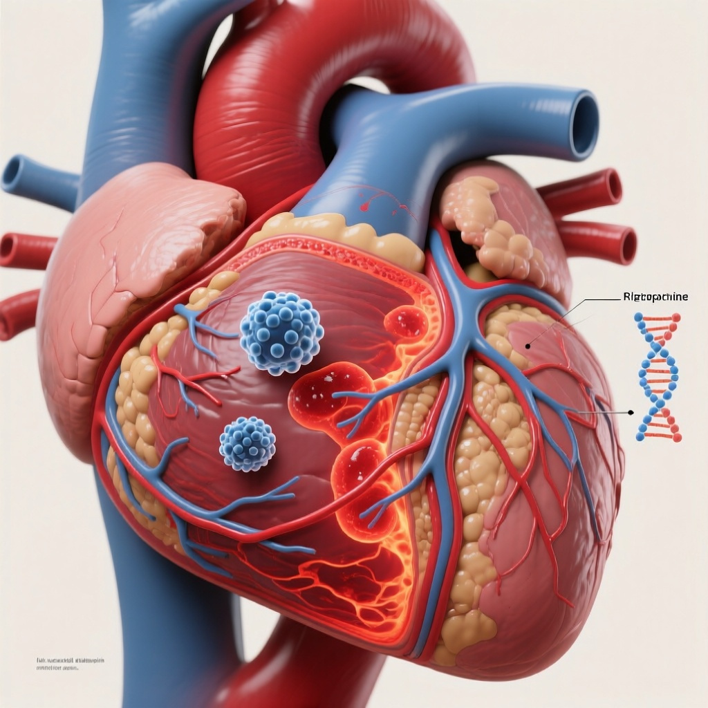
First, the study identifies circHIPK2 as a macrophage-enriched circular RNA that rises in inflammatory cardiac macrophages after myocardial infarction and tracks with the temporal inflammatory response.
Second, circHIPK2 appears to act through G3BP1-dependent stress granule formation, linking a non-coding RNA signal to macrophage inflammatory activation.
Third, macrophage-targeted inhibition of circHIPK2 in mice improved ventricular function, reduced inflammatory signaling, and attenuated post-infarction fibrosis.
Fourth, in a translational ex vivo model using living myocardial slices from patients with heart failure, circHIPK2-silenced human iPSC-derived macrophages promoted a more favorable tissue-healing response.
Clinical background and unmet need
Myocardial infarction remains a leading cause of heart failure despite major advances in reperfusion, antithrombotic therapy, secondary prevention, and systems of care. Survival after acute infarction has improved, but many patients go on to develop adverse left ventricular remodeling, progressive fibrosis, and chronic heart failure. This residual burden reflects not only myocyte loss and scar formation but also the quality, magnitude, and duration of the immune response that follows ischemic injury.
Macrophages are central to this process. In the early phase after MI, inflammatory monocyte-derived macrophages help clear necrotic debris and amplify local immune signaling. Later, reparative macrophage programs support resolution of inflammation, extracellular matrix remodeling, angiogenesis, and scar stabilization. When this transition is delayed or dysregulated, persistent inflammation can drive infarct expansion, interstitial fibrosis, chamber dilation, and systolic dysfunction.
That biology has made immune modulation an attractive therapeutic concept in ischemic heart disease, but clinical translation has been difficult. Broad anti-inflammatory strategies risk suppressing essential healing or causing systemic adverse effects, while many targets lack sufficient cell specificity. Non-coding RNAs, including microRNAs and circular RNAs, offer a potentially more selective way to reprogram disease-relevant immune cells. Circular RNAs are especially interesting because they are covalently closed, relatively stable, and increasingly implicated in gene regulation, RNA-protein interactions, and stress responses. Still, very few circRNAs have been mechanistically linked to post-MI macrophage behavior in a way that suggests therapeutic tractability.
The present study by Jung and colleagues addresses that gap by focusing on circHIPK2, a circular RNA found to be upregulated in inflammatory cardiac macrophages after MI. The investigators propose that circHIPK2 acts as a molecular switch controlling macrophage polarization and downstream cardiac remodeling.
Study design and experimental platform
This was a mechanistic and translational preclinical study that integrated in vitro macrophage experiments, a murine MI model, multimodal cardiac phenotyping, and an ex vivo human tissue platform.
In vitro, circHIPK2 expression was manipulated using siRNA-mediated knockdown and overexpression vectors. These experiments were designed to test whether changes in circHIPK2 alter macrophage inflammatory signaling, polarization state, and cytokine production.
In vivo, the authors used a mouse model of myocardial infarction combined with macrophage-targeted inhibition of circHIPK2 delivered through AAV9-mediated shRNA. Cardiac consequences were assessed with echocardiography, histology, and positron emission tomography imaging. This is an important design feature because it moves beyond simple transcriptomic association and examines organ-level consequences of immune-cell-directed intervention.
For translational relevance, the study also employed a co-culture system using living myocardial slices from patients with heart failure and human induced pluripotent stem cell-derived macrophages in which circHIPK2 had been experimentally modulated. This ex vivo approach is notable because it allows testing of immune-cell interventions in a human myocardial environment while preserving multicellular tissue architecture better than standard monolayer culture.
The primary biological endpoints were inflammatory signaling, macrophage phenotype, stress granule formation, cytokine secretion, fibrosis, and cardiac functional recovery. The abstract does not report numeric effect sizes, confidence intervals, or exact sample sizes, so interpretation must remain qualitative unless the full article is consulted.
Key mechanistic findings
circHIPK2 is induced in inflammatory cardiac macrophages after MI
The study identifies circHIPK2 as a novel circular RNA upregulated in inflammatory cardiac macrophages following myocardial infarction. Its expression pattern correlated with post-MI inflammatory dynamics, which supports the idea that this molecule is not merely a bystander biomarker but may actively participate in timing the macrophage response to injury.
From a pathobiological perspective, this is significant. The post-infarct heart is characterized by marked heterogeneity of macrophage populations, including resident and recruited subsets with overlapping but distinct functions. A circRNA enriched in inflammatory macrophages suggests a regulatory layer that may be more cell-state-specific than traditional soluble inflammatory markers.
Interaction with G3BP1 links circHIPK2 to stress granule biology
A major contribution of the paper is the reported interaction between circHIPK2 and G3BP1. G3BP1 is a well-known RNA-binding protein involved in stress granule assembly. Stress granules are dynamic ribonucleoprotein condensates that form during cellular stress and influence RNA stability, translation, and inflammatory signaling. By showing that circHIPK2 promotes stress granule formation via G3BP1, the study provides a plausible molecular bridge between non-coding RNA regulation and macrophage activation under ischemic inflammatory conditions.
This is mechanistically important because it expands the role of circRNAs beyond the commonly invoked concept of microRNA sponging. Here, circHIPK2 appears to function through RNA-protein interaction and subcellular stress-response organization. In cardiovascular immunobiology, that is a more nuanced and potentially druggable mechanism.
circHIPK2 inhibition suppresses inflammatory signaling
Silencing circHIPK2 dampened inflammatory signaling pathways and reduced secretion of pro-inflammatory cytokines. Although the abstract does not enumerate the exact cytokines or signaling nodes affected, the direction of effect is consistent with reduced inflammatory macrophage polarization. That result fits the central hypothesis that circHIPK2 acts as a molecular switch favoring a pro-inflammatory state.
If confirmed in detail, this would have therapeutic relevance because the optimal post-MI strategy is not to abolish immunity but to accelerate transition from a damaging inflammatory phase toward a reparative one. The data presented in the abstract suggest circHIPK2 inhibition may tilt macrophages in that direction.
In vivo effects on cardiac remodeling after myocardial infarction
Functional improvement in the murine MI model
In mice, macrophage-specific inhibition of circHIPK2 improved cardiac function after MI. Functional assessment included echocardiography and PET imaging, indicating that benefits were measurable not only histologically but also at the level of ventricular performance and cardiac physiology. The exact magnitude of improvement is not given in the abstract, but the direction is clinically meaningful: better ventricular function after infarction is the central goal of any remodeling intervention.
Because macrophages influence infarct healing, angiogenesis, matrix turnover, and remote myocardial inflammation, a benefit seen with cell-specific circRNA suppression supports causality more strongly than simple correlation studies. It also suggests that immune-cell-targeted RNA therapy may have effects large enough to translate into whole-organ phenotypes.
Reshaping the inflammatory milieu
The authors report that circHIPK2 inhibition reshaped the inflammatory environment. This phrase likely encompasses reduced inflammatory cytokine release, altered macrophage polarization, and possibly changes in leukocyte recruitment or persistence in the infarcted myocardium. From a translational standpoint, this matters because the inflammatory milieu after MI is not binary; it is a coordinated network involving macrophages, neutrophils, fibroblasts, endothelial cells, and cardiomyocytes. Modifying one upstream regulatory node in macrophages may therefore have systems-level consequences.
Attenuation of fibrosis progression
One of the most clinically relevant findings is that macrophage-specific circHIPK2 inhibition attenuated fibrosis progression. Post-MI fibrosis is necessary for scar stability in the infarct zone, but excessive or prolonged fibrotic remodeling contributes to stiffness, electrical heterogeneity, and heart failure progression. The therapeutic challenge has always been to preserve effective scar formation while limiting maladaptive fibrosis.
The abstract suggests that targeting circHIPK2 achieves a more favorable balance. If this reflects selective suppression of pathological inflammatory-fibrotic signaling rather than indiscriminate inhibition of wound healing, the approach would be particularly attractive. However, that distinction requires careful evaluation in the full article, including infarct rupture rates, scar quality, and timing of intervention.
Human translational relevance
The use of living myocardial slices from heart failure patients is a notable strength. These models preserve native myocardial architecture, extracellular matrix, multicellular interactions, and aspects of electrophysiology and metabolism that are often lost in conventional cell culture. By co-culturing these slices with human iPSC-derived macrophages in which circHIPK2 was silenced, the authors attempted to model how immune-cell reprogramming might affect diseased human myocardium.
According to the abstract, silencing circHIPK2 in human macrophages promoted beneficial cardiac healing in this setting, suggesting therapeutic potential even in established heart failure. That point is especially interesting because most post-MI interventions are conceptualized for the acute or subacute phase. If modulation of macrophage circHIPK2 also affects chronic inflammatory-fibrotic activity in failing myocardium, the target may have relevance beyond acute infarction.
Still, the ex vivo platform should be viewed as an intermediate translational step rather than proof of human efficacy. Myocardial slices cannot reproduce systemic immune trafficking, renal-hepatic pharmacology, neurohormonal activation, or long-term safety. Even so, they provide stronger human relevance than rodent-only data and help justify further preclinical development.
Clinical interpretation
This study sits at the intersection of cardio-immunology and RNA therapeutics. It supports a model in which macrophage behavior after MI is governed, at least in part, by circular RNA-mediated stress response programs. By identifying a circHIPK2-G3BP1-stress granule axis, the paper adds mechanistic depth and nominates a potentially targetable pathway for limiting adverse remodeling.
The work also aligns with a broader movement in cardiovascular medicine toward precision immune modulation. Prior anti-inflammatory strategies in atherosclerosis and post-MI care, including IL-1 pathway inhibition and colchicine-based approaches, have shown that inflammation matters clinically. However, those interventions are systemic. The present study advances a more cell-directed concept: modulate macrophage state directly using RNA-based tools.
Such specificity could be advantageous, especially if delivery systems can restrict activity to myeloid cells or injured myocardium. A macrophage-targeted therapy might reduce inflammatory damage while preserving systemic host defense better than broad immunosuppression. It may also avoid interfering with cardiomyocyte pathways unrelated to immune injury.
Strengths and limitations
Strengths
The study has several strengths. It combines molecular mechanism, animal physiology, imaging, tissue histology, and human translational modeling. It identifies not just an association but a candidate effector mechanism involving G3BP1 and stress granules. It uses macrophage-targeted inhibition rather than non-specific whole-heart knockdown, which sharpens biological interpretation. Finally, it addresses outcomes that matter clinically: ventricular function, inflammatory remodeling, and fibrosis.
Limitations
Several limitations should temper interpretation. First, the abstract does not provide quantitative effect sizes, variance estimates, or confidence intervals, making it impossible to judge the magnitude and precision of benefit from the summary alone. Second, all efficacy data remain preclinical. Third, AAV9-mediated shRNA is a useful experimental platform but may not represent the final clinically deployable delivery modality for transient post-MI immune reprogramming.
Fourth, macrophage polarization is more complex than a simple inflammatory-versus-reparative dichotomy. The full paper will need careful review to determine which macrophage subsets were affected and whether beneficial healing was preserved across infarct stages. Fifth, any anti-inflammatory intervention after MI raises safety questions, particularly regarding infarct healing integrity, infection risk, and off-target immune effects. Sixth, circRNA biology can be context-dependent, so validation across species, sexes, infarct sizes, and reperfusion conditions will be essential.
Finally, it remains uncertain whether circHIPK2 is best viewed as an acute post-infarction target, a chronic heart failure target, or both. Dosing window, treatment duration, and patient selection will all be major translational questions.
Implications for future research and practice
This work is unlikely to change clinical practice immediately, but it provides a strong rationale for further development of macrophage-directed RNA therapeutics in ischemic heart disease. Next steps should include detailed dose-response and timing studies, validation in large-animal MI models, biodistribution and toxicology assessment, and confirmation of human macrophage subtype effects. It will also be important to determine whether circHIPK2 inhibition interacts favorably or unfavorably with contemporary post-MI therapies such as reperfusion, beta-blockers, mineralocorticoid receptor antagonists, SGLT2 inhibitors, and anti-inflammatory agents.
Biomarker development may be another important avenue. If circHIPK2 levels in circulating monocytes, extracellular vesicles, or cardiac tissue correlate with remodeling risk, the molecule could potentially serve both as a therapeutic target and a patient-stratification marker.
For clinicians, the key message is conceptual rather than prescriptive: maladaptive post-MI inflammation may be modifiable through cell-specific RNA mechanisms, and macrophage state appears increasingly actionable as a driver of fibrosis and heart failure progression.
Conclusion
Jung and colleagues provide compelling preclinical evidence that circHIPK2 is an important regulator of macrophage inflammatory activity after myocardial infarction. Through interaction with G3BP1 and promotion of stress granule formation, circHIPK2 appears to amplify inflammatory signaling. Its macrophage-specific inhibition reduced cytokine output, improved cardiac function, and limited fibrosis in mice, while also showing promise in a human myocardial slice co-culture system.
The study is mechanistically sophisticated and translationally ambitious. Although clinical application remains distant, the findings identify a credible RNA-based immunomodulatory strategy for post-infarction remodeling and possibly chronic heart failure. In an era when residual inflammatory risk is increasingly recognized in cardiovascular disease, circHIPK2 stands out as a notable new target deserving further validation.
Funding and trial registration
The funding sources and any ClinicalTrials.gov registration number are not reported in the provided abstract. Because this is a preclinical and translational experimental study rather than a registered interventional clinical trial, a trial registration number may not apply.
References
1. Jung M, Schmidt A, Sansonetti M, Al Soodi B, Thum S, Pfanne A, Just A, Xiao K, Huang CK, Erschow S, Ricke-Hoch M, Oehlsen L, Agyapong W, Jansen K, Blume J, Büchler G, Hilbold E, Bär C, Weber N, Schinke M, Nguyen AHH, Wollert KC, Thackeray JT, Bengel F, Lachmann N, Thum T. Macrophage-specific circular RNA circHIPK2, inflammation, and fibrosis after myocardial infarction. European Heart Journal. 2026-Jun-09;47(22):2831-2847. PMID: 41667113.
2. Frangogiannis NG. The inflammatory response in myocardial injury, repair, and remodelling. Nature Reviews Cardiology. 2014;11(5):255-265.
3. Nahrendorf M, Swirski FK. Cardioimmunology: the immune system in cardiac homeostasis and disease. Nature Reviews Immunology. 2022;22(11):733-744.
4. Heusch G, Libby P, Gersh B, Yellon D, Böhm M, Lopaschuk G, Opie L. Cardiovascular remodelling in coronary artery disease and heart failure. The Lancet. 2014;383(9932):1933-1943.
5. Ibanez B, James S, Agewall S, et al. 2017 ESC Guidelines for the management of acute myocardial infarction in patients presenting with ST-segment elevation. European Heart Journal. 2018;39(2):119-177.