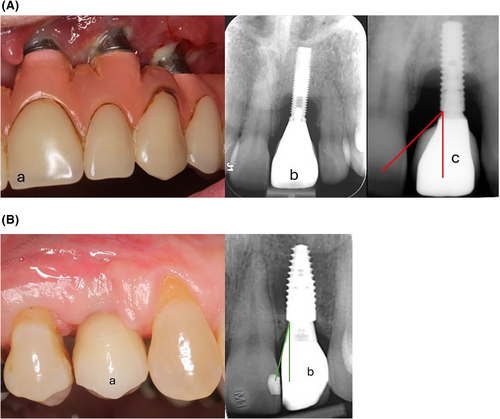
Mucosal margin mobility, thin soft-tissue phenotype, and implant connection design significantly influence the risk of peri-implantitis and peri-implant soft-tissue dehiscence (PISTD).
Platform switching and controlled prosthetic emergence angle (>45°) are important modifiable factors reducing implant complications.
Anterior implant location and daily smoking are additional risk indicators for peri-implantitis.
Peri-implantitis (8.7%) and PISTD (25%) remain prevalent complications, underscoring the need for targeted clinical interventions.
Study Background
Dental implants have become a widely accepted treatment for tooth replacement, offering improved function and aesthetics. However, implant-related complications such as peri-implantitis — an inflammatory condition causing loss of supporting bone — and peri-implant soft-tissue dehiscence (PISTD) remain significant clinical challenges that affect long-term success and patient satisfaction. Despite advances in implant technology, the multifactorial etiology of these conditions is not fully delineated, especially concerning local anatomic and prosthetic factors. Understanding modifiable risk factors during treatment planning and implant placement offers opportunities to reduce complication rates and enhance treatment outcomes.
Study Design
This investigation analyzed a cross-sectional sample of randomly selected Norwegian patients who had dental implant therapy in 2014, comprising 242 patients with a total of 732 implants. The study adopted a population-based approach and integrated clinical and radiographic assessments to capture a broad spectrum of local parameters potentially influencing implant complications.
Key data collected included:
– Soft-tissue phenotype (thin vs. thick)
– Mucosal margin mobility
– Implant connection type (external vs. internal)
– Use of platform switching
– Prosthetic emergence angle
– Implant location (anterior vs. posterior)
– Presence or absence of adjacent natural teeth
– Patient smoking habits
Mixed-effects logistic regression was employed to appropriately handle clustering of implants within individual patients and to identify independent risk indicators associated with peri-implantitis and PISTD.
Key Findings
Peri-implantitis was diagnosed in 8.7% of implants, while PISTD occurred in 25%—illustrating a substantial burden of implant-related soft and hard tissue complications.
Risk indicators for peri-implantitis included:
– Mucosal margin mobility: Implants with mobile mucosal margins showed a significantly higher risk, likely reflecting compromised soft-tissue seal and susceptibility to bacterial infiltration.
– Bone-level implant design without platform switching: Lack of platform switching correlated with increased bone loss around implants.
– Prosthetic emergence angle greater than 45°: Steeper emergence angles create unfavorable biomechanical and hygienic conditions that may promote peri-implant inflammation.
– Anterior implant location: Possibly related to anatomical and loading differences.
– Daily smoking: Smoking remains an established systemic risk factor affecting wound healing and immune response.
Risk indicators for peri-implant soft-tissue dehiscence (PISTD) were:
– Thin soft-tissue phenotype: Thin mucosa predisposes to recession and dehiscence.
– Mucosal margin mobility: As above, mobility compromises soft-tissue stability.
– Absence of adjacent teeth: Lack of neighboring teeth may reduce soft-tissue support.
– Presence of peri-implantitis: Co-existence of inflammatory bone loss exacerbates soft-tissue complications.
– External implant connection design: External connections showed higher association with PISTD compared to internal, possibly due to microgap and micro-movement effects.
These findings emphasize the interplay between prosthetic design factors and biological tissue characteristics in implant success.
Expert Commentary
This well-designed population-based study offers valuable evidence supporting previously hypothesized risk factors and highlights modifiable treatment planning elements. The use of mixed-effects modeling strengthens confidence that identified associations are independent of patient-level clustering effects.
Attention to soft-tissue phenotype is critical, suggesting potential benefits from pre-implant soft-tissue augmentation or selective implant site preparation. Preservation or restoration of adjacent teeth might also contribute to maintaining peri-implant soft-tissue integrity.
The platform switching concept, which involves using a narrower abutment on a wider implant platform to shift inflammatory infiltrate away from bone crest, showed protective effects corroborating prior biomechanical and histological studies.
Prosthetic emergence angle is easily modifiable during crown fabrication and should be optimized below 45° if possible to minimize soft-tissue stress and biofilm accumulation.
The associations noted with implant connection types and smoking reconfirm their clinical relevance, reinforcing current guidelines recommending internal connections and smoking cessation counseling.
A noted limitation is the cross-sectional design, which precludes causal inferences and temporal assessment. Also, systemic factors beyond smoking (e.g., diabetes) were not detailed. Further longitudinal and mechanistic studies are warranted to elucidate causal pathways and explore interactions between systemic and local risk factors.
Conclusion
This population-representative Norwegian study identifies several key local risk factors for peri-implantitis and peri-implant soft-tissue dehiscence, many of which are modifiable at the treatment planning stage. Optimizing soft-tissue phenotype management, selecting implant systems with platform switching and internal connections, controlling prosthetic emergence angles, and considering implant location relative to anatomical constraints are practical strategies to reduce implant complications.
Incorporating these insights into clinical protocols may improve long-term implant success rates and patient outcomes. Further research is needed to integrate local and systemic risk profiles for holistic peri-implant risk assessment and personalized treatment approaches.
Funding and Registration
The original study did not specify funding disclosures or clinicaltrials.gov registration.
References
1. Mauland EK, Bunæs DF, Lie SA, Verket A. Association Between Local Factors and Implant-Related Complications: A Population-Sampled Cross-Sectional Analysis. J Clin Periodontol. 2026; DOI: 10.1111/jcpe.13928. PMID: 42869496.
2. Schwarz F, Derks J, Monje A, Wang HL. Peri-implantitis. J Clin Periodontol. 2018;45(S20):S246-S266.
3. Linkevicius T, Apse P. Factors Influencing Peri-Implant Soft Tissue Dimensions: A Systematic Review. J Oral Rehabil. 2016;43(10):745-757.
4. Romandini M, et al. Risk Factors for Peri-Implantitis: Effect of History of Periodontitis and Smoking Habits. A Systematic Review and Meta-Analysis. J Clin Periodontol. 2020;47(3):349-368.
5. Canullo L, et al. Platform Switching and Preservation of Peri-Implant Marginal Bone. A Systematic Review and Meta-Analysis. Int J Oral Maxillofac Implants. 2017;32(3):487-498.