Highlight
– Pulmonary arterial hypertension (PAH) prognosis improves with precision myocardial strain measurement.
– Combined left ventricular global longitudinal strain (LVGLS) and right atrial reservoir strain (RARS) provide superior mortality prediction.
– This strain combination surpasses traditional cardiac magnetic resonance and clinical risk models.
– Stratification by these parameters identifies distinct 1-year mortality risk groups guiding management.
Study Background
Pulmonary arterial hypertension is a progressive, life-threatening disease characterized by elevated pulmonary vascular resistance leading to right ventricular (RV) failure. Despite advances in medical therapy, morbidity and mortality remain high. Accurate prognostic stratification is critical for tailoring treatment intensity and guiding clinical decisions. Conventional prognostic markers in PAH, including hemodynamics and cardiac magnetic resonance (CMR) metrics, have limitations in early detection and risk discrimination.
Myocardial strain parameters derived from echocardiography or CMR provide sensitive indices of myocardial deformation and function. However, optimal combinations of strain measures that best predict outcomes in PAH have not been clearly established. Given the interaction between right and left heart function, alongside atrial performance, comprehensive strain assessment is promising but understudied for prognostic utility.
Study Design
This prospective cohort study recruited 269 patients diagnosed with pulmonary arterial hypertension from June 2013 to June 2024. Participants underwent detailed myocardial strain assessment using cardiac imaging, including left ventricular global longitudinal strain (LVGLS), right ventricular global longitudinal strain (RVGLS), right ventricular free-wall longitudinal strain (RVFWLS), and right atrial phasic strain parameters.
Feature selection methods included elastic net Cox regression and stepwise Cox regression guided by the Akaike information criterion, aiming to identify the optimal combination of strain parameters for prognostic prediction. The primary endpoint was all-cause mortality, and a secondary composite endpoint included mortality and heart failure rehospitalization. Follow-up extended to a median of 39.3 months.
Key Findings
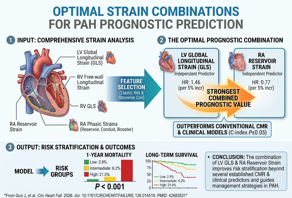
Among the 269 patients (mean age 42±15 years), 68 reached the primary endpoint and 91 reached the secondary endpoint during follow-up. The combination of left ventricular global longitudinal strain and right atrial reservoir strain emerged as the strongest predictor of prognosis.
Specifically, each 5% increment worsening in LVGLS increased mortality hazard by 46% (hazard ratio [HR] 1.46, 95% confidence interval [CI] 1.07–2.00; P=0.02), while each 5% increase in right atrial reservoir strain reduced mortality risk by 23% (HR 0.77, 95% CI 0.62–0.95; P=0.02). Both remained independent predictors when adjusted for other clinical and imaging variables.
The combined LVGLS and RARS model had significantly higher discriminatory power, as evidenced by superior C-index values compared to established CMR and clinical risk models (all P≤0.05). Patients were stratified into low-, intermediate-, and high-risk groups based on these strain parameters, demonstrating distinct 1-year mortality rates of 2.8%, 6.2%, and 21.0%, respectively (P<0.001).
Landmark analyses confirmed that this prognostic value persisted throughout long-term follow-up, underscoring its robustness.
Expert Commentary
This study importantly highlights that in PAH, incorporating both left ventricular systolic function (reflected by LVGLS) and right atrial reservoir function (RARS) provides a more comprehensive reflection of hemodynamic stress and cardiac remodeling than focusing solely on right ventricular strain. The left ventricle’s performance is increasingly recognized as crucial given the interventricular dependence and the impact of right-sided pressure overload on left-sided filling and function.
Right atrial reservoir function also reflects right heart diastolic compliance and preload conditions, offering additional prognostic insight. As noninvasive imaging techniques evolve, strain assessments are becoming more reproducible and feasible in clinical settings.
While promising, the study cohort was relatively young and single-center; results should be validated across diverse populations. The observational design limits causal inference, and the impact of strain-guided management requires future prospective trials. Integrating strain parameters with other clinical and hemodynamic markers could further refine risk scores.
Conclusion
The combined evaluation of left ventricular global longitudinal strain and right atrial reservoir strain significantly enhances prognostic stratification in pulmonary arterial hypertension beyond traditional imaging and clinical models. This dual-parameter approach identifies patient subsets with varying mortality risks, which may inform individualized clinical management and follow-up intensity.
Future studies should explore the utility of incorporating these strain parameters into PAH risk algorithms and assess whether interventions guided by strain changes improve patient outcomes. As imaging technologies advance, myocardial strain has great potential as a practical, noninvasive tool for comprehensive cardiopulmonary assessment in PAH.
Funding and ClinicalTrials.gov
Details regarding funding sources and clinical trial registration were not specified in the original publication.
References
Guo J, Leng S, He J, Yin L, Chen C, Liu X, Zhao J, Wen B, Pu S, Wan K, Wang J, Xu Y, Li Y, Han Y, Zhong L, Chen Y. Optimal Strain Combinations for Prognostic Prediction in Pulmonary Arterial Hypertension. Circulation Heart Failure. 2026 Sep 2:e014519. PMID: 42683531.
Simonneau G, Montani D, Celermajer DS, et al. Haemodynamic definitions and updated clinical classification of pulmonary hypertension. Eur Respir J. 2019;53(1):1801913.
Fine NM, Chen L, Bastiansen PM, et al. Outcome Prediction by Quantification of Right Ventricular Longitudinal Strain in Pulmonary Arterial Hypertension. Circulation Cardiovasc Imaging. 2013;6(5):554-563.