
Introduction and Context
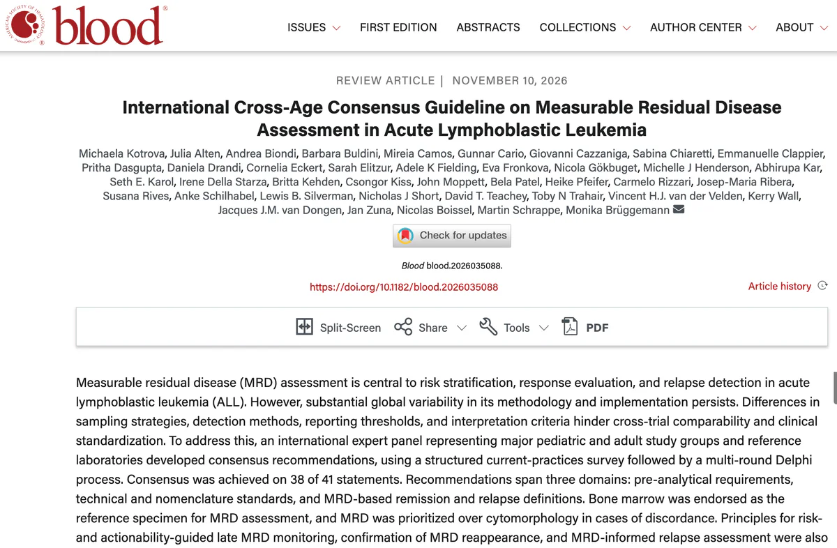
Measurable residual disease (MRD) — the small number of leukemia cells that persist after therapy — is the most powerful prognostic biomarker in acute lymphoblastic leukemia (ALL). MRD guides risk stratification, determines the depth of remission, informs decisions about therapy intensification or de‑escalation, and can signal impending relapse long before clinical signs appear. Yet, worldwide practice has been fragmented: different specimen preferences, analytic platforms (flow cytometry, PCR, next‑generation sequencing), reporting thresholds, and interpretation rules have limited cross‑trial comparability and consistent clinical use. To address these gaps, an international expert panel representing major pediatric and adult study groups and reference laboratories used a structured practice survey and a multi‑round Delphi process to produce the International Cross‑Age Consensus Guideline on Measurable Residual Disease Assessment in Acute Lymphoblastic Leukemia (Kotrova et al., Blood 2026). The panel reached consensus on 38 of 41 statements. This article summarizes the guideline’s core recommendations, what changed or was clarified from earlier practice, areas of disagreement, and practical implications for clinicians and laboratories.New Guideline Highlights
– Bone marrow is the endorsed reference specimen for MRD assessment in ALL; peripheral blood is acceptable for certain subtypes and time points (e.g., some T‑ALL or BCR::ABL1 monitoring) but is not the primary standard. – When MRD and cytomorphology disagree, MRD results take priority for clinical decision‑making. – A standardized MRD assay nomenclature was adopted to reduce reporting ambiguity across methods (flow, PCR, NGS, fusion transcript quantitation). – MRD monitoring strategy should be risk‑ and actionability‑guided: high‑risk patients and those eligible for intervention require planned late monitoring; isolated low‑level MRD may warrant confirmation before changing therapy. – Principles for confirming MRD reappearance and for MRD‑informed relapse definitions were specified to reduce false positives and unnecessary interventions. Key takeaway for clinicians: this guideline provides operational, cross‑age standards to harmonize specimen handling, technical performance, result reporting, and clinical interpretation of MRD in ALL.Updated Recommendations and Key Changes
Major advances from prior, more fragmented practice include: – Unified specimen policy: Bone marrow as the reference material replaces variable local preferences that sometimes favored peripheral blood. This is rooted in evidence that bone marrow commonly yields higher sensitivity for marrow disease. – Formalized nomenclature: The guideline prescribes how to name assays and report limits of detection, quantitation, and exact numeric results (for example, “MRDneg by flow at LOD 1 in 10,000” or “BCR::ABL1 transcript 0.01% IS by RT‑qPCR”). This replaces inconsistent lab phrases and reduces misinterpretation. – Confirmation requirement: The panel recommended confirmatory sampling and testing for low‑level or reappearing MRD before definitive clinical action, aiming to avoid premature therapy changes based on transient or false‑positive findings. – Actionability framework: MRD monitoring frequency and duration should be tailored by relapse risk, intended therapeutic options on MRD positivity (e.g., suitability for immunotherapy, transplant), and patient age. These changes reflect both accumulating technical evidence (more reliable NGS and standardized PCR approaches) and clinical experience showing that harmonized definitions improve trial comparability and patient care.Topic‑by‑Topic Recommendations
Below are the guideline’s core recommendations organized by domain. Where possible, the guideline indicates principle‑based levels (consensus vs unresolved items); the original document provides detailed technical annexes for laboratory implementation. Pre‑analytical requirements – Reference specimen: Bone marrow aspirate is the preferred and reference specimen for MRD assessment in ALL. Peripheral blood may be used for selected indications (e.g., mature B‑ALL with circulating blasts, or serial BCR::ABL1 transcript monitoring) but not as a universal substitute. – Sampling timing: MRD should be measured at pre‑specified protocol time points (end of induction, consolidation, and at risk‑guided later time points). The guideline emphasizes adherence to predefined schedules for comparability. – Sample quality: Explicit criteria for marrow sample quality (adequate cellularity, avoidance of hemodilution) are required; laboratories must document and report quality attributes with the MRD result. – Anticoagulant: No consensus was reached on a single anticoagulant for PCR‑based assays (EDTA vs heparin vs ACD), reflecting ongoing technical differences; laboratories must validate their choice and report it. Technical and nomenclature standards – Assay options: Flow cytometry (multiparameter), PCR‑based quantitative assays (fusion transcripts, clonal IG/TR rearrangement qPCR), and NGS‑based MRD assays are all accepted modalities when appropriately validated. Each must report sensitivity (limit of detection, limit of quantitation) and methodology. – Standardized reporting: Adopt the guideline’s recommended nomenclature that separates technical assay name, target, method, limit of detection/quantitation, and result (e.g., “MRDneg by 8‑color flow cytometry; LOD 0.01%).” – External quality: Participation in external proficiency and interlaboratory comparison programs is required for laboratories performing MRD testing. Reference labs should provide standardized controls and harmonized reporting templates. – Cross‑platform concordance: If MRD assays of different technologies are used longitudinally for a patient, reporting must make clear the assay change and any implications for longitudinal comparability. MRD‑based remission and relapse definitions – Remission: MRD‑negative status (below validated LOD) is defined as the absence of detectable disease by the specified assay. The guideline stopped short of mandating MRD as the exclusive definition of remission (no consensus on exclusive MRD‑only remission), but it emphasized MRD as the principal determinant when morphology is ambiguous. – Priority over morphology: When MRD and marrow morphology conflict, MRD should guide management decisions in most clinical contexts; morphology retains role in supportive diagnostic assessment. – Relapse/reappearance: Reappearance of a previously undetectable clone should be confirmed by repeat sampling and, where possible, by an orthogonal method (e.g., different PCR target or flow cytometry) before declaring molecular relapse, unless there is concurrent clinical or morphological evidence of relapse. – Action thresholds: The guideline avoids rigid universal numeric cutoffs for action because thresholds depend on disease biology, assay sensitivity, and available interventions. Instead, it recommends context‑specific action thresholds defined by clinical trial protocols or institutional consensus (for example, lower thresholds used to trigger salvage therapy when transplant or immunotherapy is planned). Monitoring strategy and late MRD surveillance – Risk‑guided frequency: High‑risk patients or those with interventions available on MRD positivity should receive more frequent and longer duration monitoring. Low‑risk patients may have less intensive surveillance. – Confirmation and trend: Isolated low‑level positive results should prompt immediate confirmatory testing and trend assessment; management should consider kinetics (rising vs stable low‑level) and patient context. – Documentation and communication: MRD reports should be delivered with clear interpretive comments for clinicians, including assay LOD, recommended follow‑up sampling, and whether results are actionable in that clinical context. Special populations and situations – Pediatrics vs adults: The guideline is cross‑age and emphasizes the same standards apply; specific monitoring frequency may differ by protocol and host factors. – Ph+ ALL (BCR::ABL1): Transcript quantification by standardized international scale (IS) methods remains an appropriate MRD method; one area without consensus was the interpretation of isolated BCR::ABL1 reconversion, which requires clinical correlation and often repeat testing. – Post‑transplant: MRD assessment post‑hematopoietic cell transplant is essential for early relapse detection; confirmed molecular reappearance often prompts preemptive interventions where available (donor lymphocyte infusion, targeted therapy).Expert Commentary and Insights
The guideline panel represented laboratory scientists, pediatric and adult clinicians, and trialists, and their deliberations illuminated both consensus and enduring controversies. Consensus perspectives – Harmonization matters: Experts agreed that standardized nomenclature, specimen selection, and reporting dramatically improve communication across centers and trials. – Confirmation before action: The requirement to confirm low‑level or reappearing MRD before changing therapy was a shared priority to avoid overtreatment and misclassification from technical artifacts. Areas of controversy and no consensus – Anticoagulant for PCR: Laboratories vary in anticoagulant choice for nucleic acid preservation; the group did not force a single standard because local validation and upstream workflows differ. This remained a technical issue for further study. – BCR::ABL1 reconversion: The panel could not agree on a single interpretation for an isolated reappearance of BCR::ABL1 transcript at low levels: some members favored immediate action in high‑risk contexts, while others favored confirmation and correlation. – Exclusive MRD definition of remission: There was insufficient consensus to declare MRD‑negativity as the exclusive definition of remission across all contexts; morphology and clinical picture retain roles in select scenarios. Expert outlook Panel members emphasized that these recommendations are practical and implementation‑oriented: they harmonize reporting and define when to act. They also highlighted the need for ongoing quality assurance, broader implementation of high‑sensitivity methods in resource‑limited settings, and prospective studies to define precise action thresholds tied to modern therapies (CAR T cells, bispecifics, targeted agents).Practical Implications
For clinicians – Request bone marrow MRD as the default for post‑induction assessment in ALL, and confirm low‑level positives before moving to salvage therapy when feasible. – Ensure MRD assay reports clearly state assay type, target, method, and limits of detection/quantitation. – Use MRD results to guide risk‑adapted therapy decisions but consider the whole clinical context, available interventions, and patient preferences. For laboratories – Adopt the standardized nomenclature, document assay performance characteristics (LOD/LOQ), participate in external quality programs, and communicate sample quality issues with clinicians. – When introducing new platforms (NGS), provide interpretive guidance about comparability to prior methods. For trialists and policy makers – Use the consensus nomenclature and definitions to facilitate pooled trials, meta‑analyses, and regulatory submissions. The guideline’s harmonized framework should improve endpoints and comparability across studies of novel therapies.Illustrative Vignette
John, a 42‑year‑old man with newly diagnosed B‑cell ALL, achieves a morphological complete remission after induction. Bone marrow MRD by 8‑color flow cytometry is negative (LOD 1 in 10,000). Under the new guideline, his treating team documents the assay name, LOD, and result, and schedules risk‑guided MRD surveillance. Six months later, a routine marrow sample shows a low‑level positive MRD signal near the assay LOD. Rather than immediate therapy escalation, the team orders confirmatory testing (repeat marrow and orthogonal PCR assay) within two weeks and discusses the potential for preemptive therapy depending on kinetics and marrow morphology. This measured approach aligns with the guideline’s emphasis on confirmation and context‑driven action.Future Directions and Research Needs
The guideline identifies several research priorities: – Prospective trials to define evidence‑based action thresholds for MRD positivity across technologies and disease subgroups. – Studies comparing anticoagulants and pre‑analytical workflows for nucleic acid‑based MRD assays to reach consensus. – Work to standardize NGS MRD reporting and cross‑platform calibration with established PCR and flow methods. – Implementation research to expand access to high‑quality MRD testing in low‑resource settings.References
– Kotrova M, Alten J, Biondi A, et al. International Cross‑Age Consensus Guideline on Measurable Residual Disease Assessment in Acute Lymphoblastic Leukemia. Blood. 2026 Nov 10. PMID: 43008231. – van Dongen JJ, Langerak AW, Brüggemann M, et al. Design and standardization of PCR primers and protocols for detection of clonal immunoglobulin and T‑cell receptor gene recombinations in suspect lymphoproliferations: report of the BIOMED‑2 Concerted Action BMH4‑CT98‑3936. Leukemia. 2003 Dec;17(12):2257–2317. – Campana D. Minimal residual disease in acute lymphoblastic leukemia. Hematology Am Soc Hematol Educ Program. 2010;2010:7–12. The 2026 cross‑age consensus guideline marks an important step toward harmonizing MRD practice in ALL. Its practical, evidence‑informed recommendations on specimen choice, reporting, confirmation of reappearance, and risk‑adapted monitoring should improve comparability across trials and help clinicians make better, more consistent decisions for patients with ALL.Specialty calculators
For clinical reference; use professional judgment.