
Highlight
- This validation study confirms the high sensitivity of GFAP and UCH-L1 plasma biomarkers in detecting intracranial injuries after mild traumatic brain injury (mTBI).
- The combined biomarker test showed high negative predictive value, supporting its use as a rule-out tool prior to CT imaging.
- Specificity and positive predictive value were low, indicating considerable false positives, especially in older patients.
- Use of the biomarker test reduced diagnostic turnaround time by more than two hours compared to CT, suggesting potential to expedite emergency department workflow.
Study Background
Mild traumatic brain injury is a common clinical issue presenting in emergency departments worldwide, accounting for a significant proportion of head trauma cases. Although most patients with mTBI have no structural brain injury, a subset harbor intracranial lesions requiring timely diagnosis and management. Head computed tomography (CT) remains the standard imaging modality to identify such injuries but entails radiation exposure, considerable cost, and logistical delays in busy emergency settings. Overuse of CT scans contributes to resource strain and increased emergency department (ED) length of stay. Emerging evidence supports the use of blood-based biomarkers as minimally invasive tools to aid diagnosis of acute brain injury. Specifically, glial fibrillary acidic protein (GFAP) and ubiquitin c-terminal hydrolase L1 (UCH-L1) have demonstrated potential in identifying intracranial lesions following mTBI by reflecting astroglial and neuronal injury, respectively. The US Food and Drug Administration (FDA) has approved a combined plasma assay for these biomarkers as an aid to detect intracranial abnormalities. However, clinical validation of this assay in US ED populations and understanding its integration with standard clinical decision rules remain necessary.Study Design
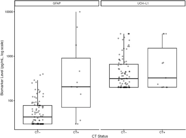
This prospective single-center validation study enrolled 104 adult patients who presented with suspected mTBI and underwent head CT at a Level I trauma center between October 2023 and June 2024. Peripheral blood samples were collected within 12 hours of injury and analyzed in the hospital clinical laboratory for GFAP and UCH-L1 plasma concentrations. Biomarker thresholds were set at GFAP >30 pg/mL and UCH-L1 >360 pg/mL to define elevated levels. The main reference standard for intracranial injury was presence of traumatic lesion detected on head CT. The study compared test sensitivity, specificity, positive predictive value (PPV), and negative predictive value (NPV) against CT findings. Additionally, the combined plasma test’s diagnostic performance was juxtaposed with the Canadian Head CT Rule, a validated clinical decision rule for mTBI. Secondary outcomes included evaluating ED length of stay and turnaround times for biomarker testing versus CT imaging.Key Findings
Among the 104 enrolled patients, 11 (approximately 10.6%) had intracranial injuries identified on CT. Biomarker analysis revealed 83 patients with elevated GFAP and/or UCH-L1 levels. The combined GFAP/UCH-L1 plasma test showed a sensitivity of 0.91 (95% confidence interval [CI]: 0.59 to 1.00), indicating high ability to correctly identify patients with intracranial injury. Specificity was low at 0.22 (95% CI: 0.14 to 0.31), driven by many false-positive biomarker elevations in patients without CT lesions. Consequently, PPV was low at 0.12 (95% CI: 0.06 to 0.21) while NPV was high at 0.95 (95% CI: 0.76 to 1.00), suggesting the assay is most useful to exclude intracranial injury. These findings align with pooled data from prior studies reporting a sensitivity of 0.97 (95% CI: 0.95 to 0.98) and a specificity of 0.28 (95% CI: 0.23 to 0.34). Notably, false-positive biomarker elevations were more frequent in older patients (median age 72 years, interquartile range [IQR] 54 to 83) compared to true negatives (median age 58.5 years, IQR 38.25 to 66). This suggests age-related factors may influence biomarker levels and warrants further investigation. A significant operational benefit was observed: median turnaround time for the GFAP/UCH-L1 test was 1.09 hours (IQR 0.87 to 1.65), over two hours faster than head CT at 3.28 hours (IQR 2.37 to 4.70). This faster result availability has implications for ED throughput and patient flow.Expert Commentary
The high sensitivity and negative predictive value affirm that plasma GFAP and UCH-L1 can function effectively as an initial screening test to rule out intracranial injury in mTBI patients. This could reduce the need for immediate CT scanning in low-risk cases, minimizing radiation exposure and resource utilization. However, the low specificity and positive predictive value demonstrate the test’s limitation in confirming intracranial injury due to frequent false positives, particularly among elderly patients. The physiological or comorbid factors contributing to elevated biomarkers in older adults remain unclear, complicating interpretation. Clinicians should therefore integrate biomarker results with clinical assessment and validated decision rules rather than relying on biomarker screening alone. Furthermore, the study’s single-center design and relatively small sample size may limit generalizability across diverse patient populations and healthcare settings. Larger, multicenter prospective trials are needed to evaluate the real-world effectiveness of incorporating biomarker testing into diagnostic protocols. These should investigate impacts on ED length of stay, cost-effectiveness, patient outcomes (short and long term), and workflow optimization. The biology underpinning GFAP and UCH-L1’s release following brain injury is well-established; GFAP is an astrocyte intermediate filament protein released with glial injury, while UCH-L1 is a neuronal cytoplasmic enzyme indicative of neuronal injury. Their combined use capitalizes on complementary injury detection mechanisms, enhancing overall test sensitivity.Conclusion
This study corroborates that the FDA-approved plasma assay measuring GFAP and UCH-L1 offers a highly sensitive method to rule out intracranial injury in mTBI patients early after injury. Integrating this blood-based biomarker test as a first-line screen before CT could decrease unnecessary imaging and reduce emergency department waiting times. However, its limited specificity, particularly in older adults, necessitates cautious interpretation and combined use with clinical criteria. Large-scale, multicenter validation is essential to determine its comprehensive effects on healthcare resource use, patient costs, and outcomes prior to widespread clinical implementation.Funding and Trial Registration
The study was conducted at a Level I trauma center with no specific funding information provided in the report. The clinical trial registration number was not stated.References
1. Papa L, Lewis LM, Falk JL, et al. Elevated levels of serum glial fibrillary acidic protein breakdown products in mild and moderate traumatic brain injury are associated with intracranial lesions and neurosurgical intervention. Ann Emerg Med. 2012;59(6):471-483.e1. 2. Yue JK, Yuh EL, Korley FK, et al. Adjusting blood-based biomarkers for detection of traumatic brain injury in emergency settings. JAMA Neurol. 2019;76(10):1135-1146. 3. Thelin EP, Zeiler FA, Ercole A, et al. Serial sampling of serum protein biomarkers for monitoring human traumatic brain injury dynamics: a systematic review. Front Neurol. 2017;8:300. 4. Canadian CT Head Rule Collaborators. The Canadian CT Head Rule for patients with minor head injury. Canadian Medical Association Journal. 2001;164(2):D65-D71.Specialty calculators
For clinical reference; use professional judgment.