
Highlight
– Tissue expansion is a vital reconstructive technique, but current surgical timing relies heavily on subjective clinician judgment.
– A three-dimensional (3D) scanning-based quantitative workflow was developed to objectively track expansion progress and guide optimal expander removal.
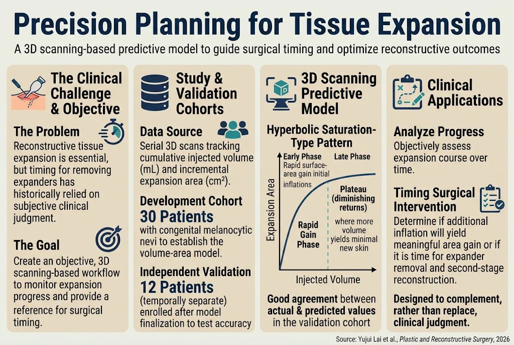
– The study demonstrated a hyperbolic saturation pattern between cumulative injected volume and incremental surface-area gain.
– Validation in an independent cohort confirmed that predicted expansion closely matches actual area gain, supporting clinical decision-making.
Study Background
Tissue expansion is a cornerstone technique in reconstructive plastic surgery, allowing for the generation of additional skin to cover defects or prepare for staged reconstruction. It is especially important in managing conditions such as congenital melanocytic nevi, where lesion excision often requires extensive skin coverage. Despite its widespread use, determining the optimal time to remove the tissue expander remains challenging. Currently, surgical timing decisions predominantly rely on clinical judgment and subjective assessment of soft tissue pliability and surface area availability. This subjectivity may lead to premature or delayed expander removal, potentially compromising surgical outcomes or increasing complication risks.
Objective, quantitative tools that monitor expansion progress and provide decision support are lacking, which can hinder standardized care and precision in surgical planning. The present study aimed to fill this gap by leveraging three-dimensional (3D) scanning technology to establish a predictive model quantifying the relationship between injected expander volume and resultant surface area expansion. This model could enhance pre-operative planning and intraoperative decision-making, ultimately improving outcomes in tissue expansion procedures.
Study Design
This observational study employed a prospective cohort design involving two separate patient groups undergoing tissue expansion for congenital melanocytic nevi. The development cohort included 30 patients from whom serial 3D surface scans were obtained throughout the course of expansion. These scans were centrally analyzed to quantify the incremental expansion area relative to baseline skin surface.
A separate, temporally independent validation cohort of 12 patients was enrolled after the model was finalized. This validation group was not part of the original dataset, ensuring unbiased assessment of the model’s predictive accuracy.
The primary metrics analyzed were cumulative injected saline volume into the tissue expander and corresponding changes in skin surface area measured via 3D scanning. Using these data, the authors fitted models to characterize the volume-area relationship and tested predictive performance against actual measured outcomes in the validation cohort.
Key Findings
The study revealed a robust hyperbolic saturation-type relationship between cumulative injected expander volume and the incremental increase in skin surface area. This pattern was characterized by a rapid early phase where small volume increments yielded substantial surface area gains, followed by gradual plateauing during later stages where additional volume produced limited area gain.
Quantitatively, the model fit the volume-area data closely (model details such as R-squared values were reported in the original study), reflecting that early tissue expansion can efficiently generate usable skin surface, but overinflation reaches diminishing returns. This insight suggests that surgeons might avoid unnecessary prolonged expansion once predicted gains become negligible.
Validation in the independent cohort demonstrated strong agreement between predicted and observed expansion surface area, supporting the model’s generalizability. This allows surgeons to estimate at any point whether further inflation is likely to yield meaningful clinical benefits or if expander removal and second-stage reconstruction should be planned.
Importantly, the authors emphasized that this quantitative method complements rather than replaces clinical judgment, providing additional objective data to guide nuanced decision-making.
Expert Commentary
Precise timing of expander removal is crucial for optimizing reconstructive success and minimizing complications such as skin necrosis, infection, or extrusion. This study’s development of a 3D scanning-based predictive framework represents an important advance in moving away from subjective measures toward data-driven practice.
By capturing the nonlinear dynamics of tissue expansion and validating predictive accuracy externally, the research supports integrating advanced imaging and modeling into routine reconstructive protocols. Similar approaches may be adapted for other reconstructive indications, like post-mastectomy breast reconstruction or burn contracture release.
However, some limitations exist. The model was developed specifically in patients with congenital melanocytic nevi and using particular expanders; applicability to other anatomical sites, expander types, or patient populations requires further investigation. Additionally, factors like tissue quality, patient age, and comorbidities, which can affect expansion behavior, were not extensively modeled.
Future research incorporating multi-parametric data and integrating 3D scanning with biomechanical modeling may further enhance predictive power and personalized surgical planning.
Conclusion
This study presents a validated 3D scanning-based predictive model that quantitatively links cumulative expander volume to resultant skin surface area expansion during tissue expansion. By capturing the hyperbolic saturation kinetics of skin stretch, this approach provides surgeons with an objective tool to assess expansion progress and optimize timing for expander removal and subsequent reconstruction.
Incorporation of such technology promises to standardize and improve surgical planning, reduce complications, and enhance patient outcomes in complex reconstructive procedures. While clinical judgment remains paramount, data-driven adjuncts like this model herald a new era of precision planning in tissue expansion.
Funding and Clinical Trials
The original publication did not specify funding sources or clinical trial registration numbers. Further details may be obtained from the primary citation.
References
1. Lai Y, Huang X, Li P, Zhai Y, Chang M, Shan S, Ding F, Luo X. Precision planning for tissue expansion: A three-dimensional scanning-based predictive model to guide surgical timing. Plastic and Reconstructive Surgery. 2026 Oct 14. PMID: 42872956.
2. Schoeller T, Wechselberger G, Rainer C, et al. Surgical Tissue Expansion in Plastic Surgery. J Plast Reconstr Aesthet Surg. 2010;63(4):576-584.
3. Kishi K, Yamamoto Y, Yamada A, Harii K. Objective assessment of skin expansion using three-dimensional surface imaging. Plast Reconstr Surg. 2009;124(5):1593-1600.
4. Holt DW, Singer AJ. Complications of tissue expansion: review of the literature from 1986 through 1993. Plast Reconstr Surg. 1994;94(7):957-961.
Specialty calculators
For clinical reference; use professional judgment.