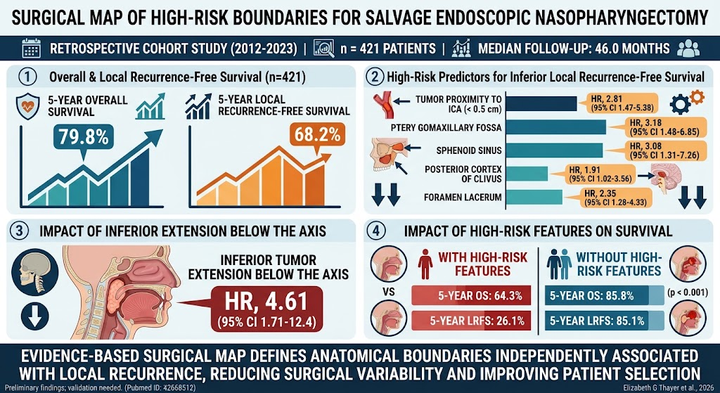
Highlight
– High-risk anatomical regions significantly impact local recurrence rates after salvage endoscopic nasopharyngectomy in recurrent nasopharyngeal carcinoma.
– Tumor proximity within 0.5 cm of the internal carotid artery, and invasion of areas like pterygomaxillary fossa and sphenoid sinus are linked to poorer outcomes.
– Patients with tumors involving these critical boundaries have substantially lower 5-year overall survival and local recurrence-free survival.
– An evidence-based surgical map has been proposed to guide resection boundaries and improve surgical decision-making.
Study Background
Nasopharyngeal carcinoma (NPC) is a malignancy arising from the epithelial lining of the nasopharynx and remains a clinical challenge, especially in the recurrent setting. Despite advances in radiotherapy and chemotherapy, local recurrence after primary treatment remains a significant obstacle to curative management. Salvage endoscopic nasopharyngectomy (SEN) has emerged as a minimally invasive surgical option offering potential for durable local control in carefully selected patients. However, the intricate anatomy of the nasopharynx and adjacent skull base structures poses surgical challenges. The lack of a clear, evidence-based anatomical framework delineating high-risk regions that influence surgical success and recurrence risks limits optimal patient selection and surgical planning.
Study Design
This retrospective cohort study reviewed 421 patients who underwent salvage endoscopic nasopharyngectomy for recurrent nasopharyngeal carcinoma between January 2012 and December 2023. Patients were assessed for tumor invasion patterns via imaging and intraoperative findings. The primary endpoints included 5-year overall survival (OS) and local recurrence-free survival (LRFS). A multivariable Cox proportional hazards model identified anatomical sites independently associated with local recurrence. These sites were integrated into a surgical map that defines critical high-risk boundaries for resection during SEN.
Key Findings
After a median follow-up of 46.0 months, the cohort demonstrated a 5-year OS of 79.8% and LRFS of 68.2%. Anatomical factors significantly associated with worse LRFS included:
– Tumor proximity less than 0.5 cm to the internal carotid artery (HR 2.813, 95% CI 1.471–5.380)
– Invasion of the pterygomaxillary fossa (HR 3.181, 95% CI 1.478–6.845)
– Sphenoid sinus involvement (HR 3.080, 95% CI 1.307–7.259)
– Posterior cortex of the clivus involvement (HR 1.906, 95% CI 1.021–3.560)
– Foramen lacerum invasion (HR 2.354, 95% CI 1.281–4.325)
– Inferior tumor extension below the axis vertebra (HR 4.611, 95% CI 1.713–12.410)
Patients harboring tumors involving these zones had substantially worse 5-year OS (64.3% vs. 85.8%, p < 0.001) and LRFS (26.1% vs. 85.1%, p < 0.001) compared to those without these high-risk features.
This suggests that tumor involvement of these anatomical landmarks should be regarded as critical risk factors and inform both the feasibility of endoscopic salvage surgery and postoperative surveillance strategies.
Expert Commentary
The authors of this study meticulously mapped anatomical risk boundaries associated with local recurrence after salvage nasopharyngectomy, helping to fill an important knowledge gap in endoscopic skull base oncology. The findings underscore the prognostic significance of tumor proximity to vital neurovascular structures like the internal carotid artery and bony landmarks such as the clivus and sphenoid sinus. By integrating these findings into a surgical guide, surgeons can better balance maximal tumor resection with safety, potentially reducing recurrence risks.
Limitations include the retrospective design and single-institution setting, which may influence generalizability. However, the large sample size and comprehensive anatomic analysis provide robustness. Prospective validation of the surgical map in diverse populations would strengthen its clinical utility.
Conclusion
This study provides an innovative, evidence-based surgical map delineating high-risk anatomical boundaries for salvage endoscopic nasopharyngectomy in recurrent nasopharyngeal carcinoma. The identification of key risk zones such as the regions adjacent to the internal carotid artery, pterygomaxillary fossa, and clivus aids in refining patient selection, planning surgical margins, and anticipating prognosis. Adoption of this surgical map into practice offers promise for improving oncologic outcomes by tailoring the surgical approach according to tumor anatomical extent. Future research should focus on prospective validation and incorporation with advanced imaging modalities to further optimize salvage strategies.
References
1. Li L, Qin Z, Guo Q, et al. A Surgical Map Defining High-Risk Anatomical Boundaries for Salvage Endoscopic Nasopharyngectomy. The Laryngoscope. 2026 Aug 26. PMID: 42655908.
2. Lee AW, Ng WT, Chan LL, et al. Management of locally recurrent nasopharyngeal carcinoma. Cancer Treat Rev. 2019;73:104–111.
3. Suh MW, Hong HJ, Kim SW, et al. Salvage endoscopic nasopharyngectomy in the treatment of recurrent nasopharyngeal carcinoma: a meta-analysis. Head Neck. 2021;43(9):2738-2747.
4. Wei WI, Sham JS. Nasopharyngeal carcinoma. Lancet. 2005;365(9476):2041-2054.