
Highlight
- Low-volume high-intensity interval training (HIIT) yields significantly higher cardiopulmonary and metabolic stress compared to time-matched moderate-intensity continuous training (MICT) in patients with coronary artery disease (CAD) during early cardiac rehabilitation (CR).
- HIIT elicited greater mean oxygen uptake (VO₂), heart rate reserve utilization, and blood lactate compared to MICT, underscoring a more potent individual physiological stimulus despite equal exercise duration.
- Participants spent substantially more time above the second ventilatory threshold and oxygen uptake reserve >90% during HIIT, correlating with higher perceived exertion ratings but supporting potential for enhanced training adaptations.
Study Background
Coronary artery disease remains a leading cause of morbidity and mortality globally, and early outpatient cardiac rehabilitation is an established intervention to improve patient outcomes by mitigating cardiovascular risk through structured exercise, education, and lifestyle modification. Effective CR exercise prescriptions aim to enhance cardiorespiratory fitness and reduce the likelihood of recurrent events. Traditionally, moderate-intensity continuous training (MICT) has been commonly prescribed. However, emerging evidence suggests that high-intensity interval training (HIIT) may elicit superior physiological adaptations in patients with CAD. Despite this, data characterizing the acute physiological responses during early CR phases—particularly comparing low-volume HIIT to time-matched MICT—remain sparse. Understanding these acute responses is crucial to optimizing individualized exercise prescriptions that balance safety with efficacy during this vulnerable phase.Study Design
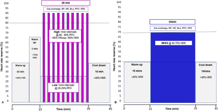
This single-center, within-subject comparison study enrolled eighteen clinically stable outpatients with CAD undergoing early cardiac rehabilitation at a UK outpatient facility. The cohort included predominantly male patients (15 male, mean age 60.1 ± 9.9 years) with preserved left ventricular ejection fraction (>35%). Each participant completed two exercise sessions, separated by at least 48 hours, under supervised conditions: 1. Low-volume HIIT protocol: 10 intervals of 1-minute cycling bouts at 85-90% of peak power output (PPO), each followed by 1-minute active recovery at 20-25% PPO, totaling 20 minutes. 2. Moderate-intensity continuous training (MICT): continuous cycling for 20 minutes at 40-70% heart rate reserve (HRR). Both sessions were preceded by a standardized 15-minute warm-up. Primary endpoints included cardiorespiratory gas exchange parameters (oxygen uptake [VO₂], carbon dioxide output [VCO₂], minute ventilation [VE]), heart rate (HR), blood pressure, rate pressure product (RPP), time spent above 90% oxygen uptake reserve (VO₂R), time relative to ventilatory thresholds 1 and 2 (VT1/VT2), blood lactate concentration (BLa), energy expenditure, and rating of perceived exertion (RPE).Key Findings
The comparative analysis revealed that low-volume HIIT induced markedly greater physiological stress across multiple domains compared to time-matched MICT: – Mean oxygen uptake (VO₂) and oxygen uptake reserve (%VO₂R) were significantly higher during HIIT (P < 0.003), reflecting augmented aerobic demand. – Time spent above 90% VO₂R and above ventilatory threshold 2 (VT2) was substantially increased during HIIT (approximately 834 ± 387 seconds vs. 393 ± 465 seconds, P < 0.0004), consistent with intense metabolic challenge. – Heart rate reserve utilization (%HRR) and rate pressure product (RPP) were elevated, indicating heightened cardiac workload. – Blood lactate concentrations were significantly greater following HIIT, indicative of enhanced anaerobic glycolytic contribution. – Participants reported higher perceived exertion during HIIT sessions (RPE 13 ± 1 vs. 11 ± 1, P < 0.003), although sessions were time-matched. Energy expenditure was similar between protocols, indicating the difference in intensity rather than total work volume accounted for the physiological divergence. These data confirm that even low-volume HIIT protocols can provoke greater acute ventilatory, hemodynamic, and metabolic demands than traditional continuous training in early-stage CR patients.Expert Commentary
The study by Begg et al. substantiates the growing consensus that adequately dosed HIIT can safely be employed early in cardiac rehabilitation to maximize physiological stimulus. The increased time above critical ventilatory thresholds and oxygen uptake reserve exemplifies the superior intensity-related stimulus essential for inducing central and peripheral cardiovascular adaptations. Elevated RPP during HIIT merits clinical attention but remains within tolerable limits in stable CAD patients with reasonable left ventricular function. While higher perceptual effort might pose challenges for widespread adherence, personalized coaching and patient education can mitigate this barrier. The findings align with current European Society of Cardiology guidelines endorsing HIIT as an effective cardiac rehabilitation modality, underpinning its incorporation into individualized exercise programs. Limitations include the small sample size and predominance of male patients, potentially limiting generalizability, and the acute nature of the study precludes direct inference about long-term training benefits or safety outcomes. Future research should focus on larger, diverse cohorts with longitudinal follow-up assessing clinical endpoints and quality of life metrics associated with early HIIT implementation.Conclusions
This within-subject comparison unequivocally demonstrates that low-volume HIIT delivers a more potent physiological and metabolic stimulus compared to time-matched MICT in early outpatient cardiac rehabilitation patients with CAD. These findings inform clinical decision-making by supporting the incorporation of personalized HIIT protocols to optimize cardiorespiratory fitness improvements during a critical window of secondary prevention. Careful patient selection and monitoring remain essential to balance efficacy with safety.Funding and ClinicalTrials.gov
The reported study does not specify funding sources or clinical trial registration information.References
1. Begg B, Griffiths TD, Drane AL, et al. Physiological responses during high intensity interval training and moderate intensity continuous training in patients attending early cardiac rehabilitation. Arch Phys Med Rehabil. 2026 Nov 11. PMID: 43014437. 2. European Society of Cardiology (ESC) Guidelines on Cardiovascular Disease Prevention in Clinical Practice, 2021. 3. Weston M, et al. High-intensity interval training in patients with lifestyle-induced cardiometabolic disease: a systematic review and meta-analysis. Br J Sports Med. 2014;48(16):1227-1234. 4. Rognmo Ø, et al. Cardiovascular risk of high- versus moderate-intensity aerobic exercise in coronary heart disease patients. Circulation. 2012;126(12):1436-1440.Specialty calculators
For clinical reference; use professional judgment.