
Study Background
Primary aldosteronism (PA) represents the most common form of secondary hypertension characterized by autonomous aldosterone production, frequently caused by unilateral adrenal adenomas or bilateral adrenal hyperplasia. Precise subtyping—distinguishing between unilateral (lateralized) and bilateral disease—is critical as it guides treatment strategy, with unilateral cases potentially curable by adrenalectomy. Currently, adrenal venous sampling (AVS) is the gold standard for PA subtype diagnosis but is invasive, technically challenging, and not widely available in all centers. Furthermore, PA can coexist with mild autonomous cortisol secretion (MACS), complicating clinical assessment and influencing prognosis and management. Noninvasive biomarkers that can reliably identify PA lateralization and coexisting cortisol excess remain an unmet clinical need.Study Design
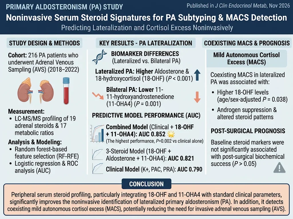
This prospective cohort study included 216 patients diagnosed with primary aldosteronism who underwent AVS between 2018 and 2022. The investigation aimed to evaluate peripheral serum steroid profiles for their diagnostic value in PA lateralization and detection of MACS, and their potential in predicting postsurgical biochemical outcomes. Serum steroid profiling was performed using liquid chromatography-tandem mass spectrometry (LC-MS/MS), quantifying 19 adrenal steroids and calculating 17 metabolic ratios reflecting adrenal steroidogenesis pathways. Feature selection employed random forest-based recursive feature elimination to identify the most predictive steroid markers. Logistic regression models were developed and validated to predict lateralization status and postoperative biochemical success, with performance assessed by area under the receiver operating characteristic curve (AUC).Key Findings
The study identified distinct steroid signatures differentiating lateralized PA from bilateral PA. Notably, lateralized PA patients exhibited significantly elevated serum aldosterone and 18-hydroxycortisol (18-OHF) levels and decreased 11-hydroxyandrostenedione (11-OHA4) levels compared with bilateral cases (all P-values <0.01). A parsimonious three-steroid model incorporating 18-OHF, aldosterone, and 11-OHA4 demonstrated robust discrimination between unilateral and bilateral disease with an AUC of 0.821, comparable to a clinical model based on serum potassium, plasma aldosterone concentration (PAC), and plasma renin activity (PRA) (AUC 0.790, P=0.351). Integrating these steroid markers with clinical parameters yielded a combined model with superior diagnostic accuracy (AUC 0.852), significantly outperforming the clinical model alone (P=0.002). This suggests that adding steroid profiling enhances noninvasive lateralization detection beyond traditional hormonal measures. Regarding coexisting MACS, patients with lateralized PA plus cortisol excess exhibited higher 18-OHF concentrations (age/sex-adjusted P=0.038), along with androgen suppression and altered steroid patterns indicative of altered adrenal steroidogenic activity. However, baseline steroid profiles did not predict postoperative biochemical cure, indicating that while steroid signatures aid diagnosis and subtype classification, their prognostic utility post-adrenalectomy warrants further study.Expert Commentary
This study elegantly demonstrates that peripheral serum steroid profiling, utilizing advanced LC-MS/MS techniques, can serve as a noninvasive, physiologically informative adjunct to AVS in PA subtyping. Elevated 18-OHF—a metabolite primarily produced in the zona fasciculata—may reflect lateralized adrenal pathology with enhanced steroidogenic output beyond aldosterone. Concurrent androgen suppression and steroidogenic shifts in patients with MACS highlight the complex interplay between aldosterone and cortisol pathways in this population. The combined steroid and clinical parameter model addresses critical limitations of AVS, including invasiveness, cost, and technical demand, potentially expanding access to confident subtype diagnosis. Nevertheless, it is essential to recognize that steroid profiling currently cannot fully replace AVS, particularly in complex cases or centers where AVS expertise is available. Larger multicenter studies are needed to validate these findings across diverse populations and to explore longitudinal steroid profile changes in relation to treatment outcomes.Conclusion
Serum steroid profiling represents a promising, noninvasive tool to improve the diagnosis and subtyping of primary aldosteronism, effectively identifying lateralized disease and concomitant mild autonomous cortisol secretion. Addition of steroid biomarkers to conventional clinical measures enhances diagnostic accuracy and may guide personalized management strategies. However, the prognostic role of baseline steroid signatures for predicting biochemical cure post-adrenalectomy remains unclear and requires further investigation. This approach holds potential to complement or, in select cases, reduce dependence on adrenal venous sampling, facilitating earlier and more precise therapeutic decision-making in PA.Funding and ClinicalTrials.gov
The study credentials indicate support by institutional and possibly public research grants; however, no specific funding disclosures or clinical trial registration numbers were provided in the source publication.References
1. Park SS, Noh J, Jung S, et al. Noninvasive serum steroid signatures of lateralization and coexisting cortisol excess in primary aldosteronism. J Clin Endocrinol Metab. 2026 Nov 10; PMID: 43004931. 2. Funder JW, Carey RM, Mantero F, et al. The management of primary aldosteronism: case detection, diagnosis, and treatment: an Endocrine Society clinical practice guideline. J Clin Endocrinol Metab. 2016;101(5):1889-1916. 3. Mulatero P, Monticone S, Bertello C, et al. Genetic and clinical characterization of primary aldosteronism. J Clin Endocrinol Metab. 2015;100(12):E1694-E1704.Specialty calculators
For clinical reference; use professional judgment.