Logical section structure
1. Title
2. Highlights
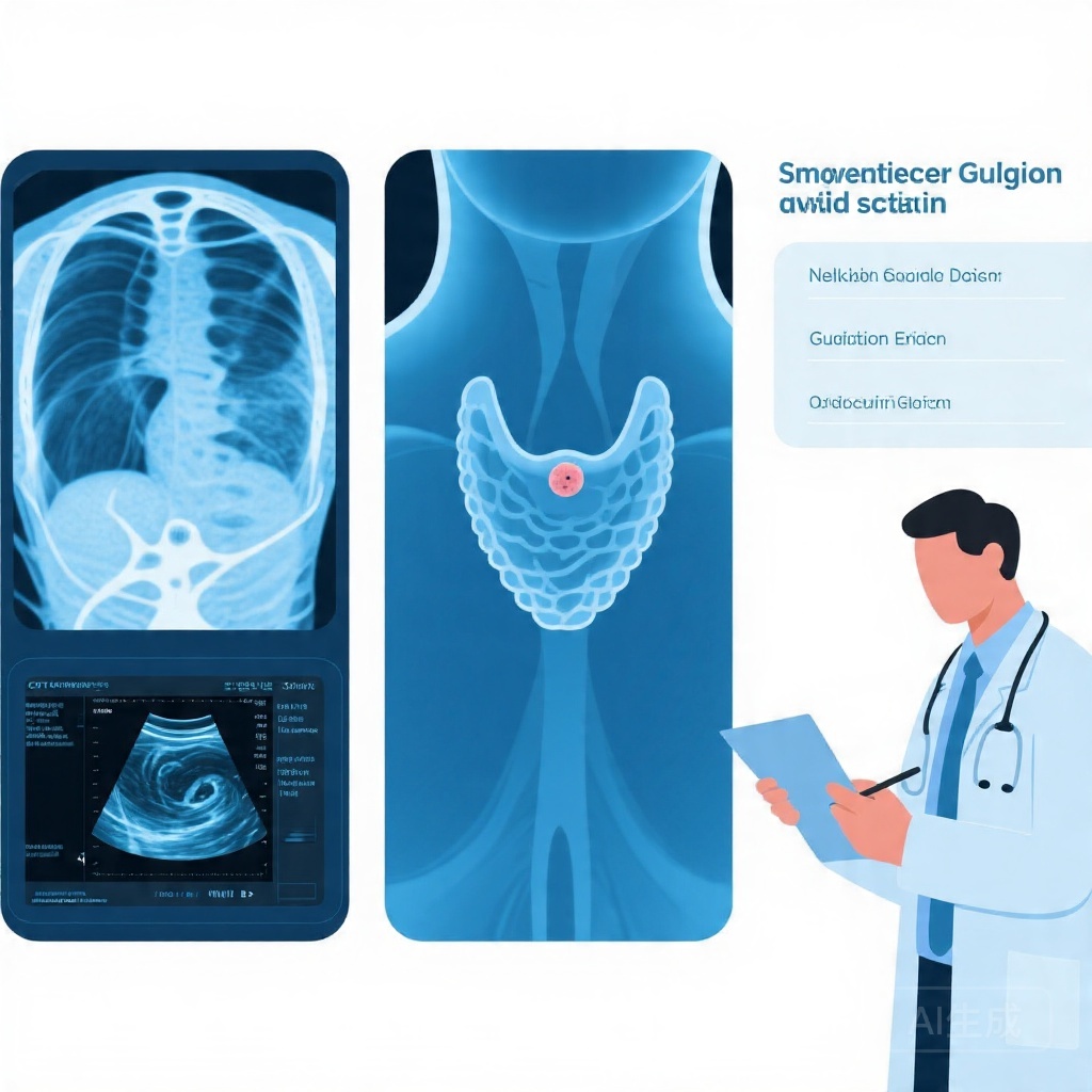
3. Clinical background and why thyroid incidentalomas are challenging
4. Study design and guideline comparison framework
5. Key findings
6. Interpretation and expert commentary
7. Clinical implications and conclusion
8. Funding and clinical trial registration
9. References
Title
Restrictive Thyroid Incidentaloma Guidelines Cut Workload Dramatically but Miss a Small Number of Low-Risk Cancers
Highlight
Thyroid incidentalomas found on imaging for unrelated reasons are common, but most conventional imaging-detected lesions have a very low malignancy risk.
In this retrospective cohort, nuclear imaging-detected incidentalomas carried a much higher cancer rate than lesions found on ultrasound, CT, or MRI.
Simulation of guideline strategies showed that more restrictive approaches could avoid most evaluations, but they would miss a small number of cancers with favorable prognosis.
Background
Thyroid incidentalomas are thyroid nodules discovered incidentally during imaging performed for non-thyroid indications. As cross-sectional imaging and nuclear medicine studies have become more widely used, incidental thyroid findings have increased as well. This creates a recurring clinical dilemma: should every incidental lesion be evaluated, or should testing be limited to cases most likely to harbor clinically meaningful malignancy?
The answer matters because thyroid cancer detection is not uniformly beneficial. Intensive workup can identify occult malignancies, but it also increases ultrasound referrals, fine-needle aspiration procedures, patient anxiety, and the risk of overdiagnosis of indolent disease. On the other hand, overly restrictive criteria may reduce burden but allow some malignancies to go unrecognized. Existing guidelines attempt to balance these competing harms, yet they differ substantially in thresholds for evaluation.
This study directly addresses that variability by examining real-world outcomes in a large referral cohort and by simulating how different guideline pathways would alter diagnostic workload and cancer detection.
Study design
This retrospective cohort study included all patients referred to Zuyd Thyroid Center between 2018 and 2023 for thyroid nodule evaluation. The investigators identified patients with incidentalomas and collected data on diagnostic outcomes, treatment, and complications.
A separate guideline-based simulation analysis compared three approaches to incidental thyroid lesion evaluation:
The European Thyroid Association (ETA) guideline, described here as the most intensive strategy.
The American Thyroid Association (ATA) approach, including a scenario without clinical warning signs.
The Dutch Federation of Medical Specialists guideline, described as the most restrictive strategy.
The main outcomes were malignancy detection and diagnostic workload, defined by the proportion of incidentalomas that would trigger further evaluation under each guideline strategy.
Key findings
Among 1825 referred patients, 630 had one or more incidentalomas, representing 34.5% of the cohort. The median age was 66 years, with an interquartile range of 54 to 74 years, and 70.6% were female. This demographic profile is typical of thyroid nodule referral populations, which often skew toward older adults and women.
A key result was the large difference in malignancy rate according to the imaging modality that first detected the incidentaloma. Nuclear imaging-detected incidentalomas had a malignancy rate of 21.1%, whereas conventional imaging-detected incidentalomas had a malignancy rate of only 1.8% (p < 0.001). This gap is clinically important because it suggests that the pretest probability of cancer varies substantially by detection context.
In the simulation analysis, the ETA guideline recommended evaluation of all cases. Unsurprisingly, this strategy identified all malignancies, but at the cost of the highest diagnostic workload.
The ATA strategy, when applied without clinical warning signs, would have evaluated 85.3% of patients and still identified all malignancies. In practical terms, this means that a modest degree of selectivity can reduce unnecessary assessments without losing detected cancers in this cohort.
The Dutch guideline was far more restrictive. It would have avoided evaluation in 96.9% of patients, which dramatically reduced workload, but 7 malignancies among 528 unevaluated cases would have been missed, corresponding to 1.3% of those not evaluated. Importantly, all of these missed cancers were subtypes with good prognosis even if left undetected, suggesting that the clinical harm of delayed diagnosis may be limited in this context.
The study therefore highlights a central tradeoff in thyroid incidentaloma management: detecting every malignancy is not necessarily equivalent to improving outcomes, particularly when many detected cancers are indolent.
Interpretation and expert commentary
The most practice-relevant message from this study is that “one-size-fits-all” evaluation of thyroid incidentalomas is difficult to justify for conventional imaging findings. The observed malignancy rate of 1.8% in conventional imaging-detected incidentalomas is low, and the number needed to evaluate to find one malignancy is therefore high. This is exactly the setting in which overdiagnosis becomes a major concern.
By contrast, incidentalomas detected on nuclear imaging appear to represent a biologically and clinically different group. The much higher malignancy rate of 21.1% supports more aggressive evaluation in that subgroup. This distinction is important for clinicians who may otherwise apply a uniform “incidentaloma” label across very different imaging contexts.
The simulation results also help explain why guideline differences persist. A more inclusive strategy maximizes sensitivity, but it may overwhelm services and expose many patients to low-value testing. A restrictive strategy improves efficiency, but at the risk of missing some cancers. The key question is not simply whether a malignancy is found, but whether finding it changes outcomes enough to justify the downstream cascade of testing and treatment.
Several limitations should temper interpretation. First, this is a retrospective single-center referral cohort, so the findings may not generalize to all imaging populations or healthcare systems. Referral bias is likely: patients sent to a thyroid center are not the same as all patients with incidental nodules on imaging. Second, the simulation analysis estimates guideline impact rather than measuring prospective outcomes under each guideline. Third, the study does not fully resolve whether the missed cancers under the restrictive strategy would have become clinically significant over time, although the reported favorable subtypes suggest relatively low immediate risk.
Even with these caveats, the study aligns with the broader movement in thyroid cancer care toward risk-adapted management and avoidance of overdiagnosis. In practice, incidentaloma management should consider the imaging modality, nodule size, patient age, comorbidity, life expectancy, and the presence of suspicious clinical features rather than relying solely on detection itself.
Clinical implications and conclusion
This study supports a more nuanced approach to thyroid incidentalomas. Conventional imaging-detected lesions generally confer a low malignancy yield, meaning that broad evaluation strategies may generate substantial workload for relatively little diagnostic gain. More restrictive criteria can sharply reduce unnecessary workup, though clinicians must accept the possibility of missing a small number of low-risk cancers.
The challenge for future guideline development is to improve selection criteria so that clinically relevant malignancies are detected while indolent disease and unnecessary procedures are minimized. Better risk stratification tools, including imaging features, clinical context, and potentially molecular or artificial intelligence-based approaches, may help narrow this gap.
For now, the study provides a strong reminder that incidental does not mean clinically equivalent. How a thyroid lesion is found matters, and management should reflect that context.
Funding and clinical trial registration
The abstract provided does not report funding or clinical trial registration information.
References
1. Willems JIA, van Twist DJL, Pickwell KM, van Battum PLH, Bianchi R, Sluiter T, Medici M, Peeters RP, Tummers-de Lind van Wijngaarden RFA. Diagnostic Yield of Thyroid Incidentaloma Evaluation and the Impact of Guideline Variability: Insights from a Retrospective Cohort Study. Thyroid. 2026 Apr 14:10507256261440405. PMID: 41980912.
2. Haugen BR, Alexander EK, Bible KC, et al. 2015 American Thyroid Association Management Guidelines for Adult Patients with Thyroid Nodules and Differentiated Thyroid Cancer. Thyroid. 2016;26(1):1-133.
3. Tessler FN, Middleton WD, Grant EG, et al. ACR Thyroid Imaging, Reporting and Data System (TI-RADS): White Paper of the ACR TI-RADS Committee. J Am Coll Radiol. 2017;14(5):587-595.
4. Hoang JK, Langer JE, Middleton WD, et al. Managing Incidental Thyroid Nodules Detected on Imaging: White Paper of the ACR Incidental Thyroid Findings Committee. J Am Coll Radiol. 2015;12(2):143-150.
5. Brito JP, Morris JC, Montori VM. Thyroid Cancer: Zealous Imaging Has Increased Detection and Treatment of Low-Risk Tumors. Endocrinol Metab Clin North Am. 2014;43(2):469-488.