
Highlights
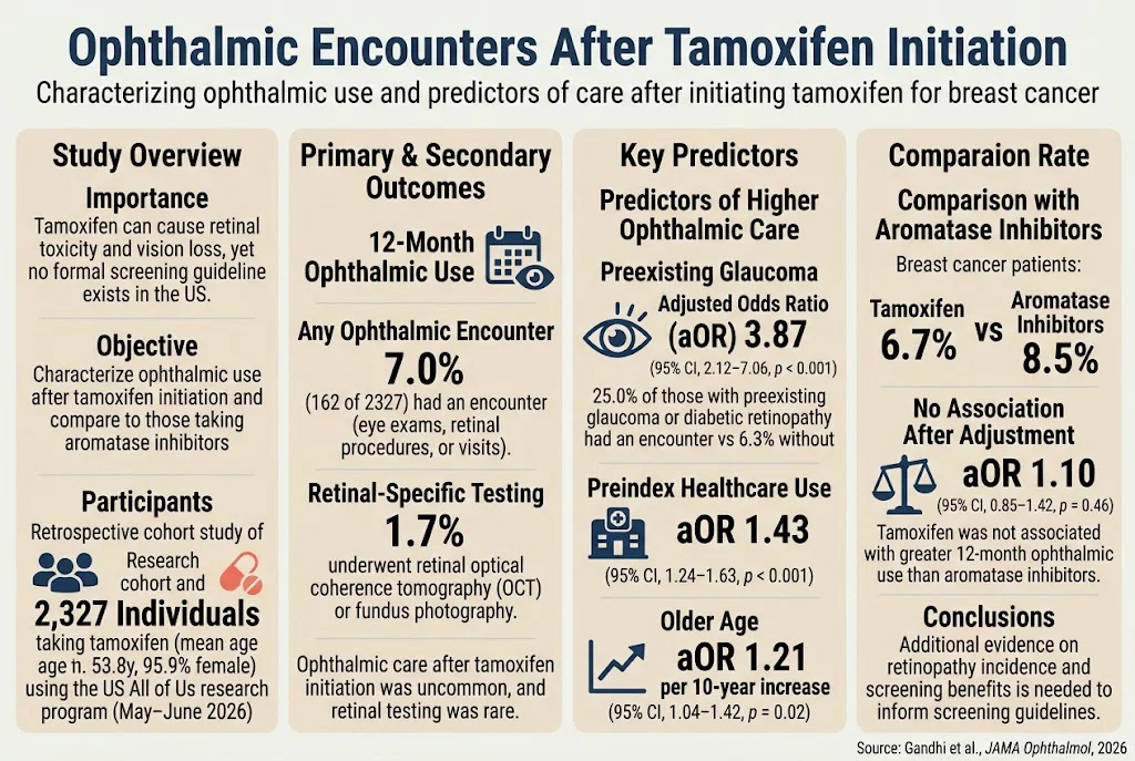
- Ophthalmic encounters within the first year after initiating tamoxifen are uncommon (7.0%), with retinal-specific testing (OCT or fundus photography) even rarer (1.7%).
- Preexisting ocular conditions (glaucoma, diabetic retinopathy), older age, and higher prior healthcare use increase likelihood of ophthalmic visits post-tamoxifen.
- Comparative analysis finds no significant increase in ophthalmic use within 12 months between tamoxifen and aromatase inhibitor users after adjustment for confounders.
- Advanced retinal imaging reveals early outer retinal changes (notably ellipsoid zone reflectivity loss) preceding overt retinopathy, offering a potential early biomarker.
- Current US practice lacks standardized screening guidelines for tamoxifen-induced retinal toxicity, highlighting a crucial evidence gap for timely detection and management.
Background
Tamoxifen, a selective estrogen receptor modulator, is widely used as adjuvant hormonal therapy in hormone receptor-positive breast cancer. Despite its efficacy, tamoxifen carries risks of ocular toxicity, notably tamoxifen retinopathy characterized by crystalline deposits and outer retinal damage, which can progress to vision loss. However, unlike hydroxychloroquine retinopathy, tamoxifen-related retinal toxicity has less clearly defined epidemiology, natural history, and screening protocols, especially in the US where formal guidelines are lacking. Ophthalmic surveillance after tamoxifen initiation remains heterogeneous, with variable clinical practices and uncertainty regarding incidence rates and risk stratification. The advent of multimodal retinal imaging technologies such as optical coherence tomography (OCT) and ultra-widefield imaging allows for more sensitive detection of subtle structural retinal changes, potentially enabling earlier diagnosis.Key Content
Retrospective Cohort Evidence on Ophthalmic Use
A recent large retrospective cohort study using the All of Us dataset (Gandhi et al., 2026) evaluated ophthalmic encounters within 12 months following first tamoxifen prescription in 2,327 adults (mean age 53.8 years, predominantly female). Only 7.0% had any ophthalmic encounter, and retinal-specific testing with OCT or fundus photography was performed in merely 1.7%. Predictors of higher ophthalmic utilization included preexisting glaucoma (adjusted odds ratio [aOR] 3.87), diabetic retinopathy, higher overall healthcare use before index date, and older age. In a comparator subgroup with breast cancer taking aromatase inhibitors (n=2,809), 8.5% had an ophthalmic encounter versus 6.7% in tamoxifen users. After controlling for confounders, tamoxifen was not associated with increased likelihood of ophthalmic visits (aOR 1.10). These findings indicate that ophthalmic evaluation post-tamoxifen initiation is uncommon and comparable to that seen with aromatase inhibitor therapy, suggesting screening or referral is not routinely triggered by tamoxifen use. However, patients with preexisting ocular comorbidities merit closer monitoring.Imaging and Structural Biomarker Studies
Emerging prospective imaging studies provide mechanistic and early detection insights. A cohort study (Ponzini et al., 2026) employing swept-source OCT quantified reflectivity changes in the ellipsoid zone (EZ), external limiting membrane (ELM), and retinal pigment epithelium (RPE) among tamoxifen-treated women without overt retinopathy. Significant reductions in EZ reflectivity at specific foveal and perifoveal locations preceded clinical retinopathy detection, while ELM and RPE reflectivity remained stable. These subtle EZ disruptions may serve as an early biomarker for retinal involvement, potentially guiding closer surveillance or therapeutic adjustments. Another study (Toklu et al., 2023) reported increased choroidal thickness and decreased choroidal vascularity index in tamoxifen users compared to controls, suggesting early subclinical chorioretinal vascular alterations. These imaging biomarkers underscore the potential to identify patients at risk well before irreversible retinal damage occurs, although longitudinal studies are needed.Prevalence and Epidemiological Insights
Epidemiologic data remain limited. A Canadian cohort found a low prevalence (1.6%) of definitive tamoxifen retinopathy using multimodal and ultra-widefield imaging, with peripheral retinal changes not significantly more frequent in tamoxifen users than controls, consistent with a low incidence of manifest retinopathy in routine care (Karp et al., 2025). Pediatric cases, although extremely rare, highlight that high cumulative doses can produce crystalline retinopathy even in children, warranting early ophthalmic screening in such populations (Samara et al., 2023).Comparisons with Hydroxychloroquine-Associated Retinopathy
Hydroxychloroquine (HCQ) retinopathy shares similarities but is better characterized with established screening guidelines. HCQ retinopathy risk is influenced by cumulative dose, duration, kidney disease, and tamoxifen co-use, with tamoxifen posing an additional retinopathy risk factor (Melles and Marmor, 2024; Matsuoka et al., 2026). Studies on HCQ emphasize the importance of early detection through multimodal imaging and annual screening after 5 years of use. The risk factors and structural changes identified for tamoxifen retinopathy suggest that lessons from HCQ monitoring may inform tamoxifen-related clinical practice.Other Ocular Effects and Risk Factors
Beyond retinopathy, tamoxifen has been associated with corneal thickness reduction and subtle visual field changes, which may have implications for glaucoma risk, warranting ophthalmologic glaucoma screening in tamoxifen recipients with preexisting risk factors (Dogan et al., 2025). Furthermore, tamoxifen use carries thromboembolic risks including rare retinal artery occlusion, highlighting the need for comprehensive ocular and systemic risk assessment (Liu et al., 2025).Current Screening Practices and Guideline Gaps
A population-based Korean study (Kim et al., 2024) revealed that only 6.5% of tamoxifen users underwent baseline ophthalmic screening within 1 year, and subsequent monitoring was performed in less than 30% of these patients, with fundus photography dominating as the assessment modality and OCT underutilized. These findings reflect a lack of standardized screening protocols and variable adherence. In the US, no formal tamoxifen retinal toxicity screening guideline exists despite recognized toxicity risks. This contrasts with established screening paradigms for HCQ.Expert Commentary
The accumulated evidence indicates that while tamoxifen retinal toxicity can cause irreversible vision loss, ophthalmic surveillance post-treatment initiation is infrequent and unsystematic. The low utilization of retinal OCT and fundus photography suggests missed opportunities for early detection of subclinical retinal changes. Risk stratification incorporating preexisting ocular comorbidities (glaucoma, diabetic retinopathy), age, and cumulative tamoxifen exposure may help identify patients who would benefit from enhanced monitoring. The emerging imaging biomarkers, particularly ellipsoid zone reflectivity loss and choroidal structural changes, offer promising tools for early, noninvasive detection before clinical retinopathy manifests. From a mechanistic standpoint, tamoxifen’s retinal toxicity may involve photoreceptor and retinal pigment epithelial damage, associated with choroidal vascular alterations, paralleling some features of macular telangiectasia type 2 and other Müller cell-involving retinopathies. However, the current literature underscores significant gaps: there is a paucity of longitudinal data on tamoxifen retinopathy incidence, natural history, and impact on visual function. Equally, clinical trial data addressing the benefit-risk ratio of routine retinal screening remain absent. Integration of these insights with clinical guidelines could improve patient safety by instituting tailored screening algorithms. The balance between surveillance burden and early detection benefit remains to be defined.Conclusion
Tamoxifen poses a known but poorly characterized risk of retinal toxicity. Ophthalmic reviews and retinal-specific monitoring within the first year of tamoxifen therapy are uncommon, even among patients with breast cancer. Preexisting glaucoma and diabetic retinopathy significantly increase ophthalmic service use post-tamoxifen initiation. Retinal changes identified by advanced imaging suggest early subclinical involvement preceding overt retinopathy. Current lack of formal screening guidelines in the US leads to heterogeneity in clinical practice. Further research is urgently needed to delineate the incidence and progression of tamoxifen retinopathy, validate early imaging biomarkers, and assess the clinical utility of screening interventions. This evidence will inform consensus guidelines, optimize patient care, and mitigate vision loss risks.References
- Gandhi S, Balas M, Wang P, McKay BR, Kertes PJ, Martens R. Ophthalmic Encounters After Tamoxifen Initiation. JAMA Ophthalmol. 2026 Oct 29; doi:10.1001/jamaophthalmol.2026.4646. PMID: 42955022.
- Ponzini F, Rossi S, Balestrazzi A, et al. Early detection of tamoxifen-induced retinal injury: ellipsoid zone reflectivity as a sensitive structural marker. BMC Ophthalmol. 2026 Jun 18;26(1):351. doi:10.1186/s12886-026-05042-x. PMID: 42316354.
- Karp CL, Tzekov R, Goldstein SM, et al. Prevalence and spectrum of manifestations of tamoxifen retinopathy as assessed by multimodal and ultra-widefield retinal imaging. Can J Ophthalmol. 2025 Jun;60(3):186-191. doi:10.1016/j.jcjo.2024.09.005. PMID: 39433277.
- Dogan A, Fidan I, Horozoglu F. Influence of tamoxifen on ocular morphology and function. Oncologist. 2025 May 8;30(5):oyaf103. doi:10.1093/oncolo/oyaf103. PMID: 40377437.
- Kim YJ, Lee JH, Cho AR, et al. Nationwide Screening Practices for Tamoxifen Retinal Toxicity in South Korea: A Population-Based Cohort Study. J Clin Med. 2024 Apr 9;13(8):2167. doi:10.3390/jcm13082167. PMID: 38673439.
- Samara WA, Hong NK, Tham Y-C. Crystalline Retinopathy in a 6-Year-Old Boy with History of High-Dose Tamoxifen Use. Retin Cases Brief Rep. 2023 Nov 1;17(6):751-753. doi:10.1097/ICB.0000000000001280. PMID: 35383718.
- Toklu Y, Acar M, Celik T, et al. Tamoxifen related chorioretinal structural changes. Cutan Ocul Toxicol. 2023 Sep;42(3):109-117. doi:10.1080/15569527.2023.2220388. PMID: 37272809.
- Melles RB, Marmor MF. The risk of toxic retinopathy in patients on hydroxychloroquine therapy: A systematic review and meta-analysis. JAMA Ophthalmol. 2024;142(1):103-111. doi:10.1001/jamaophthalmol.2023.5479. PMID: 36705365.
- Liu Y, Sun S, Kuang Z, et al. Real-world assessment of thromboembolic risk associated with tamoxifen. Sci Rep. 2025 Jul 30;15(1):27820. doi:10.1038/s41598-025-13585-0. PMID: 40739320.
Specialty calculators
For clinical reference; use professional judgment.