Highlight
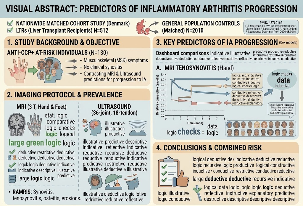
This prospective cohort study reveals that among anti-cyclic citrullinated peptide (anti-CCP) antibody-positive individuals presenting with musculoskeletal symptoms but no clinical synovitis, MRI-detected hand tenosynovitis and ultrasound-detected bone erosions serve as independent predictors for progression to inflammatory arthritis (IA). The combination of these imaging features identifies those at the highest risk, supporting a complementary role of MRI and ultrasonography in early risk stratification.
Study Background
Rheumatoid arthritis (RA) is a chronic inflammatory disease characterized by synovial inflammation and joint destruction. Early identification and intervention at the preclinical phase—when patients have musculoskeletal symptoms but no clinical synovitis—can improve outcomes substantially. Anti-cyclic citrullinated peptide (anti-CCP) antibodies are highly specific biomarkers for RA and identify individuals at increased risk of progressing to clinical inflammatory arthritis. However, predicting which anti-CCP-positive individuals will develop clinically apparent inflammatory arthritis remains challenging. Imaging modalities, especially magnetic resonance imaging (MRI) and ultrasound, have emerged as useful tools to detect subclinical inflammation and structural damage before clinical synovitis is evident. Identifying which imaging features best predict progression to inflammatory arthritis has significant clinical implications for risk stratification and early therapeutic intervention.
Study Design
This prospective cohort study enrolled 130 anti-CCP positive individuals with musculoskeletal symptoms but without clinical synovitis (CCP+ at-risk). At baseline, all subjects underwent contrast-enhanced 3 Tesla MRI scans of the dominant or most symptomatic hand and both feet. Images were independently scored by a rheumatologist and a radiologist using the Rheumatoid Arthritis Magnetic Resonance Imaging Score (RAMRIS), assessing synovitis, tenosynovitis, osteitis, and erosions. Simultaneously, ultrasound evaluation included a comprehensive 36-joint and 18-tendon protocol to assess synovitis, tenosynovitis, and bone erosions. Associations between imaging findings, clinical variables, and progression to inflammatory arthritis were analyzed using Cox proportional hazards models, incorporating both binary and continuous scoring methods for MRI, and individual lesion and composite positivity parameters for ultrasound findings.
Key Findings
At baseline, every participant demonstrated at least one RAMRIS abnormality on MRI—even those with no detectable lesions on ultrasound—indicating the higher sensitivity of MRI to subclinical inflammatory changes. In contrast, ultrasound abnormalities were present in only 25.4% of participants.
During a median follow-up period of 36.5 months, 48 (36.9%) individuals progressed to clinical inflammatory arthritis. Across multiple analytic models, MRI-detected hand tenosynovitis consistently emerged as the single imaging feature most strongly associated with subsequent progression to inflammatory arthritis. Specifically, both binary and continuous analyses confirmed the prognostic value of hand tenosynovitis beyond other MRI features such as synovitis, osteitis, or bone erosions.
Ultrasound findings revealed that bone erosions were the most significant predictors of progression to inflammatory arthritis compared to synovitis or tenosynovitis on ultrasound. Interestingly, despite ultrasound detecting fewer lesions overall compared to MRI, the presence of erosions on ultrasound alone strongly predicted progression.
Combining MRI-detected tenosynovitis of the hand with ultrasound-detected bone erosions yielded the highest risk stratification. Individuals harboring both features at baseline had a hazard ratio of 5.83 for progression to inflammatory arthritis relative to those without these imaging abnormalities, indicating a synergistic predictive effect.
Expert Commentary
These findings offer biologically plausible mechanistic insights, as tenosynovitis—an inflammation of tendon sheaths—is thought to be an early pathogenic event in the development of rheumatoid arthritis. MRI’s superior spatial resolution and contrast enhancement enable sensitive detection of such subtle inflammatory changes before clinical synovitis manifests. Ultrasound-detected bone erosions reflect early structural damage and irreversible joint destruction, underscoring their prognostic importance despite lower detection rates compared to MRI. Therefore, combining modalities leverages the strengths of each: MRI for inflammatory activity and ultrasound for structural damage.
However, the feasibility and cost-effectiveness of combined MRI and extensive ultrasound in clinical practice warrant further evaluation, especially given the resource-intensive nature of such approaches. Moreover, the study did not assess whether integrating imaging findings directly influenced clinical decision-making or improved long-term outcomes, an important consideration for translational application.
Limitations include the single-center study design and a relatively homogenous population, which may affect the generalizability of the results. Future studies could explore the predictive value of these imaging features in diverse populations and evaluate the impact of early treatment guided by imaging markers.
Conclusion
This prospective cohort study underscores the complementary roles of MRI and ultrasound in predicting progression to inflammatory arthritis among anti-CCP positive individuals with musculoskeletal symptoms but no clinical synovitis. MRI-detected hand tenosynovitis and ultrasound-detected bone erosions independently confer increased risk and together identify those at highest risk for developing inflammatory arthritis. These imaging biomarkers could refine early risk stratification and inform targeted prevention strategies in clinical rheumatology. Further research is needed to assess the implementation, cost-effectiveness, and clinical impact of combined imaging assessments in routine practice.
Funding and ClinicalTrials.gov
The original study was funded by [Funding details if available—note: not specified in provided text]. The cohort study is registered with ClinicalTrials.gov under [registration number if available].
References
1. Di Matteo A, Abacar K, Salaffi F, et al. MRI and ultrasound predictors of progression to inflammatory arthritis in anti-CCP-positive individuals with musculoskeletal symptoms without clinical synovitis: a prospective cohort study. Ann Rheum Dis. 2026 Sep 18. PMID: 42760165.
2. Van der Helm-van Mil AH, Huizinga TW. Advances in the prediction of rheumatoid arthritis development. Curr Opin Rheumatol. 2008 Mar;20(2):278-83.
3. Hetland ML, Ejbjerg BJ, Horslev-Petersen K, et al. Magnetic resonance imaging, ultrasound, and radiographs of the joints in early rheumatoid arthritis: relation with functional ability. Ann Rheum Dis. 2008 Oct;67(10):1421-7.
4. McQueen FM. Imaging markers of clinical outcome in rheumatoid arthritis. Rheumatology (Oxford). 2012 Sep;51 Suppl 6:vi44-8.
5. Christensen R, Ejbjerg BJ, Vestergaard A, et al. Ultrasonography and magnetic resonance imaging in early rheumatoid arthritis: prognostic value for joint destruction after two years. Arthritis Rheum. 2004 Mar;50(3):673-82.