Title
Corticostriatal Connectivity Reorganizes Before and After Motor Decline in Parkinson Disease
Highlights
• In Parkinson disease, changes in nigrostriatal dopamine loss were linked to distinct patterns of corticostriatal functional reorganization.
• Hyperconnectivity between the posterior caudate and primary motor regions emerged early, before about 50% putaminal dopamine depletion, and disappeared later as caudate dopamine also declined.
• In contrast, the posterior putamen showed loss of connectivity with posterior cortical regions around the same depletion threshold, aligning with faster motor deterioration.
• The findings support a staged network model in which different striatal subregions contribute differently to prodromal and symptomatic Parkinson disease.
Background
Parkinson disease (PD) is classically defined by degeneration of nigrostriatal dopaminergic neurons, leading to dopamine depletion in the striatum and downstream dysfunction of basal ganglia-thalamocortical circuits. This process disrupts the balance between direct and indirect pathway signaling, ultimately producing bradykinesia, rigidity, and gait impairment. Although the dopaminergic deficit is a core biological feature of PD, the clinical expression of motor symptoms depends not only on the degree of dopamine loss but also on how brain networks adapt to that loss over time.
A major unanswered question has been whether corticostriatal functional connectivity (FC) changes in a uniform way as nigrostriatal degeneration advances, or whether different striatal territories show distinct adaptive and maladaptive patterns. This distinction matters clinically because the striatum is functionally heterogeneous: the caudate and putamen participate in partially different loops, and these loops may be reorganized differently across prodromal disease, early PD, and more established motor impairment. Understanding these changes may help explain why some patients remain relatively stable despite substantial dopaminergic loss, whereas others show rapid motor deterioration once a threshold of degeneration is crossed.
The present study addresses this unmet need by integrating dopamine transporter PET with resting-state functional MRI to characterize how corticostriatal networks change across a broad range of nigrostriatal degeneration. The authors also included patients with idiopathic REM sleep behavior disorder (iRBD), a recognized prodromal synucleinopathy, to help anchor the degeneration continuum before overt PD.
Study Design
This was a cross-sectional multimodal neuroimaging study conducted in a large clinical cohort. Investigators recruited 326 patients with Parkinson disease and 29 patients with idiopathic REM sleep behavior disorder, all of whom underwent brain resting-state functional MRI, N-(3-[18F]fluoropropyl)-2β-carbomethoxy-3β-(4-iodophenyl) nortropane PET, and Unified Parkinson’s Disease Rating Scale assessment. In addition, 40 healthy controls were used to define the extent of striatal dopamine depletion in the PD-spectrum cohort, and another 40 healthy controls were used to compare corticostriatal FC with the patient group.
The study focused on seed-based FC in the anterior and posterior caudate and putamen on both the more affected and less affected sides. Using a sliding window method, the authors evaluated FC changes as mean putaminal dopamine declined from approximately 70% to 20%. This approach allowed them to examine network changes across a continuum rather than treating PD as a single static state. The primary clinical anchor was motor symptom severity, with attention to how FC alterations related to progression of motor deterioration.
Key Findings
The most important result was that the posterior caudate and posterior putamen did not behave similarly as dopamine depletion worsened. Instead, they showed temporally distinct patterns of connectivity change that likely reflect different stages of network compensation and failure.
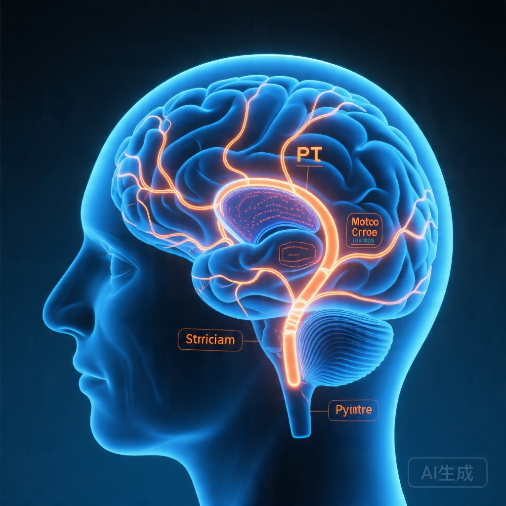
On the more affected side, the posterior caudate demonstrated elevated FC with the primary motor cortex and paracentral lobule. This hyperconnectivity was already present before approximately 50% putaminal dopamine depletion, peaked around that level, and then disappeared when caudate dopamine itself became abnormally reduced. In practical terms, this suggests that early in the disease course, when dopaminergic loss is still evolving, the motor cortex and posterior caudate may engage in a compensatory or reorganization response that temporarily preserves function. However, this response does not persist indefinitely.
By contrast, the more affected posterior putamen showed reduced FC with the superior parietal cortex, precuneus, and cuneus when putaminal dopamine depletion reached approximately 50%. After this point, motor symptoms worsened more rapidly and appeared to progress linearly. This pattern is particularly important because the putamen is strongly tied to sensorimotor processing. Loss of its connectivity with posterior cortical regions may therefore mark a transition from relatively compensated network dysfunction to overt clinical decline.
Notably, the posterior putamen did not show the same early hyperconnectivity with motor cortex that was observed in the posterior caudate. Instead, its FC remained relatively unchanged with motor cortex while its broader connections with posterior cortical hubs declined. This dissociation supports the idea that the caudate and putamen are not simply interchangeable striatal nodes but participate in different compensatory and degenerative trajectories.
Clinically, the authors interpret these findings as evidence of a functional reorganization of corticostriatal circuitry across the course of PD. The posterior caudate appears to participate in an early adaptive phase associated with slower motor progression, whereas posterior putaminal disconnection aligns more closely with the onset of motor symptoms and accelerated deterioration. This staged pattern may help explain why motor impairment in PD often seems to “tip” once a certain burden of dopaminergic loss has accumulated.
Although the abstract does not report detailed effect sizes, confidence intervals, or p values, the sample size was substantial for a multimodal imaging study, and the use of dopamine transporter PET strengthens the biological interpretation by linking FC changes to a measurable marker of nigrostriatal degeneration rather than to clinical diagnosis alone.
Clinical and Translational Interpretation
These findings reinforce a modern view of Parkinson disease as a network disorder as much as a neurotransmitter deficiency state. Dopamine depletion remains central, but the brain’s response to that depletion determines how clinical dysfunction emerges. The study suggests that functional connectivity changes may first reflect compensation, then reorganization, and eventually failure of the motor network.
For clinicians, the main translational insight is that motor symptom severity may not map linearly to dopamine depletion. Patients can exhibit substantial dopaminergic loss while maintaining relatively stable motor performance, likely because some circuits are still compensating. Once posterior putaminal connectivity with posterior cortical regions begins to decline, however, the system may lose this reserve, and motor progression may accelerate.
This framework could eventually help in several areas. First, imaging biomarkers might be used to distinguish prodromal from early symptomatic stages more precisely than clinical examination alone. Second, connectivity patterns may improve patient stratification in neuroprotective or disease-modifying trials. Third, the data may inform future interventions that aim not only to replace dopamine but also to preserve or restore network-level function.
Expert Commentary
The study has several strengths. It combines PET and resting-state MRI in a relatively large PD cohort, includes prodromal iRBD participants, and examines degeneration as a continuum rather than as a binary diagnosis. That design is well suited to capturing nonlinear network responses. The seed-based analysis of anatomically defined striatal territories also aligns with known basal ganglia functional organization.
At the same time, the study has important limitations. Its cross-sectional design cannot prove that the observed connectivity changes cause motor progression; they may instead be biomarkers of parallel disease processes. Resting-state FC is also sensitive to motion, medication state, preprocessing choices, and physiological noise, all of which can influence results in movement disorders. The abstract does not specify whether patients were scanned in a defined medication state, which is relevant because dopaminergic therapy can alter both symptoms and connectivity. In addition, the study focuses primarily on motor symptoms and does not address cognitive or psychiatric manifestations that may also relate to corticostriatal reorganization.
Generalizability is another consideration. Although the cohort is sizable, it comes from a specific research environment, and imaging-based thresholds such as “about 50% putaminal dopamine depletion” may not transfer directly to individual patients or other cohorts. Finally, the work is mechanistically persuasive but still observational; longitudinal studies are needed to determine whether posterior caudate hyperconnectivity truly predicts preservation, compensation, or later decline.
Even with these caveats, the biological narrative is compelling. The posterior caudate may support a temporary bridge during prodromal and early PD, while the posterior putamen may represent the critical node whose disconnection heralds clinically meaningful motor worsening. This distinction adds nuance to the long-standing view that PD progression is driven simply by “more dopamine loss equals worse symptoms.”
Conclusion
This study shows that corticostriatal connectivity in Parkinson disease is dynamically reorganized as nigrostriatal degeneration advances. Early posterior caudate hyperconnectivity with motor cortex may reflect a compensatory phase, whereas later posterior putaminal disconnection from posterior cortical regions appears more closely tied to the transition into faster motor decline. Together, these findings support a staged network model of PD and highlight the value of combining dopaminergic PET with functional MRI to understand disease progression. The work strengthens the case for connectivity-based biomarkers, but longitudinal validation will be essential before these measures can guide routine clinical care.
Funding and clinicaltrials.gov
The abstract provided does not list funding sources or a clinicaltrials.gov registration number. No trial registration was reported in the supplied material.
References
1. Yoo HS, Kim HK, Park M, Ahn SJ, Lee JH, Ryu YH, Lyoo CH. Functional Reorganization of Corticostriatal Connectivity Across the Degree of Nigrostriatal Degeneration in Parkinson Disease. Neurology. 2026-04-17;106(10):e214812. PMID: 41996661.
2. Poston KL, Eidelberg D. Functional brain networks and abnormal connectivity in Parkinson disease. J Neurol Neurosurg Psychiatry. 2009;80(2):197-204.
3. Wu T, Hallett M. The basal ganglia and neural networks in tremor, bradykinesia, and rigidity. Handb Clin Neurol. 2018;160:135-147.
4. Schapira AHV, Chaudhuri KR, Jenner P. Non-motor features of Parkinson disease. Nat Rev Neurosci. 2017;18(7):435-448.