Proposed Section Structure
This article is organized around the clinical problem, the study’s multimodal design, the regional biology of cardiac sarcoidosis lesions, the newly described humoral autoimmune axis, and the implications for diagnosis and treatment. The major sections are Highlights, Clinical Background, Study Design and Methods, Key Results, Mechanistic and Clinical Interpretation, Limitations, and Conclusion, followed by Funding/Registration and References.
Highlights
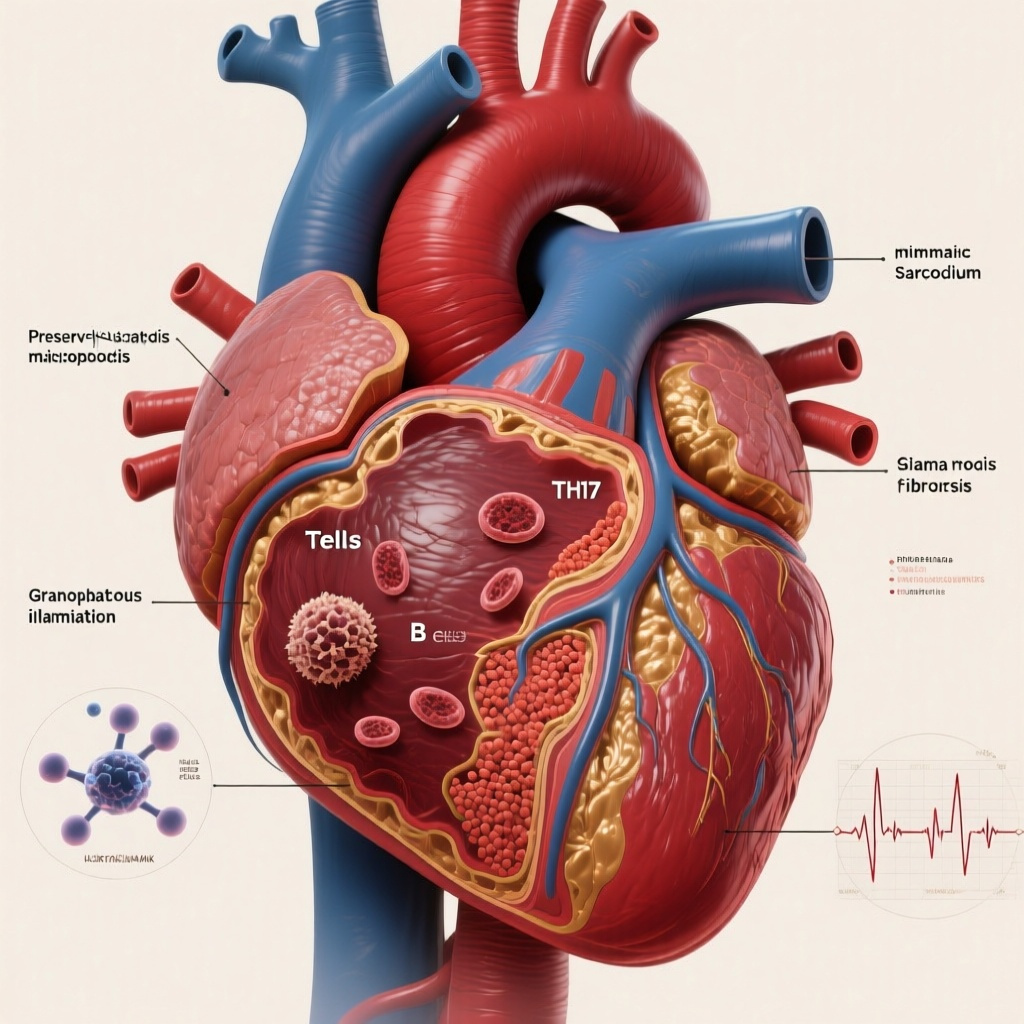
First, this study suggests that cardiac sarcoidosis is not a single static lesion but a spatially evolving disease with distinct molecular programs in preserved myocardium, granulomatous tissue, and fibrosis.
Second, cardiomyocytes and fibroblasts in apparently preserved regions expressed chemotactic and inflammatory programs, while granulomatous regions were enriched for activated macrophages and Th17-skewed T cells.
Third, fibrotic regions contained tertiary lymphoid structures with clonally expanded B cells and plasma cells, supporting an intracardiac humoral immune response rather than nonspecific bystander inflammation.
Fourth, reconstructed antibodies from expanded B-cell clones recognized periplakin (PPL), a desmosomal protein, and other cardiac-expressed peptides, raising the possibility that autoantigen-directed immunity contributes to arrhythmia and progressive myocardial injury in cardiac sarcoidosis.
Clinical Background
Cardiac sarcoidosis (CS) remains one of the most clinically consequential and biologically enigmatic inflammatory heart diseases. It can present with ventricular arrhythmias, conduction disease, sudden cardiac death, and progressive heart failure. Diagnosis is often difficult because myocardial involvement is patchy, histology can miss active lesions, and imaging findings may overlap with those of myocarditis or arrhythmogenic cardiomyopathies. Even when CS is recognized, key mechanistic questions persist: why granulomas form in some myocardial areas but not others, why fibrosis often persists without active granuloma, and what drives ongoing electrical instability and ventricular dysfunction.
Historically, sarcoidosis has been viewed primarily as a T-cell/macrophage-mediated granulomatous disease. That framework explains some aspects of pathology but leaves major gaps. In particular, it does not fully account for the patterned coexistence of normal-appearing myocardium, granulomatous inflammation, and scar, nor does it explain why some patients continue to worsen despite conventional immunosuppression. The current study addresses these gaps using single-cell and spatial transcriptomics, coupled with antibody reconstruction from clonally expanded cardiac B cells.
Study Design and Methods
Neyazi and colleagues performed integrative molecular profiling of explanted or biopsied human CS cardiac tissue, analyzing preserved, granulomatous, and fibrotic regions separately. The approach combined single-cell transcriptomics, spatial transcriptomics, immune repertoire analysis, and functional characterization of antibodies derived from clonally expanded B cells.
The study design is especially notable for its spatial resolution. Rather than treating the diseased heart as biologically homogeneous, the investigators mapped gene expression to histopathologic compartments. This design is highly relevant in CS, where the patchwork distribution of lesions is central to both diagnosis and clinical behavior.
A key methodological feature was the unexpected identification of expanded cardiac B-cell clones containing rearranged immunoglobulin sequences. The authors reconstructed antibodies from these clones and screened them against libraries spanning the human peptidome as well as microbial and allergen peptides to identify candidate antigens.
The abstract does not report conventional effect estimates such as hazard ratios or confidence intervals, which is typical for discovery-oriented translational work rather than interventional clinical trials. The major endpoints were mechanistic and descriptive: cell-type composition, compartment-specific transcriptional programs, clonal B-cell expansion, and antigen reactivity of reconstructed antibodies.
Key Results
1. Cardiac sarcoidosis shows region-specific immune and stromal programs
The study found substantial differences in both cellular composition and gene expression across preserved, granulomatous, and fibrotic myocardial regions. This reinforces the concept that CS is a spatially staged disorder rather than a uniform inflammatory cardiomyopathy.
In preserved myocardium, cardiomyocytes and fibroblasts were not biologically quiescent. Instead, they activated chemotactic and inflammatory signaling programs that could recruit and sustain myeloid and lymphoid infiltrates. This is an important observation because it implies that tissue appearing relatively intact by histology may still be molecularly diseased and capable of propagating inflammation.
2. Cardiomyocytes exhibited arrhythmogenic and inflammasome-associated signatures
Cardiomyocytes upregulated transcripts linked to arrhythmogenicity and inflammasome activation, including signals associated with pyroptosis, a lytic inflammatory form of programmed cell death. This finding may help connect pathology to phenotype. In CS, electrical instability often appears disproportionate to the visible burden of inflammation, and this study suggests that stressed cardiomyocytes themselves may participate in creating a pro-arrhythmic substrate.
From a translational standpoint, this is highly relevant. It implies that arrhythmia risk in CS may reflect not only scar architecture but also ongoing inflammatory reprogramming of surviving myocytes.
3. Granulomas were enriched for fusion-competent macrophages and Th17-skewed T cells
Within granulomatous regions, macrophages expressed modulators of cell-cell fusion, a biologically plausible feature given the multinucleated giant cells seen in granulomas. The T-cell compartment showed Th17 skewing, and these T cells upregulated B-cell-activating factor, implicating crosstalk between cellular and humoral immunity.
This observation broadens the conventional model of CS. Rather than a purely macrophage/T-cell granulomatous process, the data support an immune network in which Th17-biased inflammation can foster B-cell survival, maturation, and antibody production.
4. Fibrotic regions contained tertiary lymphoid structures
One of the most striking findings was the presence of tertiary lymphoid structures in fibrotic, nongranulomatous regions. These ectopic lymphoid aggregates contained mature B cells and plasma cells with evidence of clonal expansion. Such structures are increasingly recognized in chronic autoimmune and inflammatory disorders as local sites of antigen presentation, affinity maturation, and sustained antibody generation.
In clinical terms, this may explain why fibrosis in CS is not simply an inactive scar. Fibrotic myocardium may remain immunologically active, functioning as a niche for ongoing adaptive immune responses even after granulomas have waned.
5. Reconstructed antibodies recognized cardiac autoantigens, including periplakin
The most provocative result was that antibodies reconstructed from expanded intracardiac B-cell clones did not react to microbial or allergen peptide libraries but instead recognized periplakin (PPL), a desmosome-associated protein, along with other peptides expressed on cardiac cells. This argues against a simple infectious or environmental mimic in the assayed systems and supports true autoreactivity.
Periplakin is of particular interest because it is linked to desmosomal integrity. The authors propose that PPL autoreactivity may help explain phenotypic overlap between CS and arrhythmic desmosomal cardiomyopathies, including ventricular arrhythmias and progressive myocardial dysfunction. If validated, this could represent a mechanistic bridge between immune-mediated myocardial injury and desmosome-related electrical disease.
Mechanistic and Clinical Interpretation
This study proposes a coherent disease model with three interconnected compartments. In preserved myocardium, stressed cardiomyocytes and activated fibroblasts generate chemoattractant signals. In granulomatous zones, macrophage fusion programs and Th17-dominant inflammation amplify cellular immunity. In fibrotic regions, tertiary lymphoid structures sustain B-cell maturation and autoantibody production. Together, these findings suggest that CS progresses through overlapping inflammatory and autoimmune circuits rather than through isolated episodes of granuloma formation.
The identification of a heart-resident humoral autoimmune axis has several potential clinical implications. First, it may partially explain incomplete responses to corticosteroid-centered therapy, which is often effective but inconsistent. Second, it raises the possibility that selected patients could benefit from therapies targeting B cells, plasma cells, or cytokine pathways that support B-cell survival, such as BAFF-related signaling. Third, it suggests that autoantibodies or autoantigen-specific immune signatures might eventually serve as biomarkers for diagnosis, disease activity, arrhythmic risk, or treatment response.
These findings also intersect with current challenges in electrophysiology. In CS, sudden death and ventricular tachyarrhythmias may occur even when imaging suggests only limited active inflammation. Autoimmune targeting of desmosomal or junctional proteins could plausibly destabilize electrical coupling, intensifying risk beyond what scar burden alone predicts.
Strengths and Limitations
The major strengths of the study are its use of human tissue, its high-resolution multimodal profiling, and its integration of spatial biology with functional antibody reconstruction. The discovery pipeline is especially compelling because it moved beyond descriptive immunophenotyping to antigen identification.
Several limitations deserve emphasis. First, this is a mechanistic study, not a therapeutic or prognostic trial, so the immediate clinical applicability remains inferential. Second, the sample size is likely limited, as is common in studies of human CS tissue, especially explanted hearts. Third, the identification of PPL as a candidate autoantigen is exciting but requires validation across larger and independent cohorts, along with testing of specificity versus other inflammatory or arrhythmogenic cardiomyopathies. Fourth, absence of reactivity to the screened microbial libraries does not definitively exclude infectious or molecular mimicry mechanisms outside the assayed epitopes.
Finally, it remains uncertain whether anti-PPL responses are pathogenic drivers, epiphenomena of tissue injury, or both. Establishing causality will require orthogonal approaches, including serologic studies, animal or cellular models, and correlation with clinical phenotypes such as ventricular arrhythmias, conduction disease, and response to immunomodulation.
Practice and Research Implications
For clinicians, the study reinforces that CS should not be conceptualized solely as granulomatous myocarditis. Preserved-appearing myocardium and fibrotic scar may both harbor active pathogenic programs. This has implications for how clinicians interpret imaging, biopsy sampling, and disease progression.
For investigators, the work opens several avenues: validation of PPL and other candidate autoantigens; development of blood-based or tissue-based biomarker assays; exploration of B-cell-directed treatment strategies; and comparison of autoimmune signatures across CS, giant cell myocarditis, lymphocytic myocarditis, and arrhythmogenic cardiomyopathy.
For translational cardiology more broadly, the study illustrates how single-cell and spatial methods can reveal disease mechanisms invisible to conventional histology. Similar analytic frameworks may help define autoantigens and tissue-resident immune circuits in other poorly understood inflammatory heart diseases.
Conclusion
Neyazi and colleagues provide one of the most mechanistically integrated views of cardiac sarcoidosis to date. Their data support a model in which preserved myocardium, granulomatous lesions, and fibrosis each contribute distinct inflammatory functions, culminating in a localized autoimmune B-cell response within the heart. The identification of periplakin as a candidate autoantigen is particularly notable because it offers a plausible link between immune injury and arrhythmogenic structural heart disease. Although the findings are not yet practice-changing, they materially advance the field and point toward more precise immunophenotyping, biomarker development, and potentially B-cell-focused therapeutic strategies in CS.
Funding and ClinicalTrials.gov
The abstract does not report a ClinicalTrials.gov registration number, which is expected for this mechanistic human tissue study rather than a registered interventional trial. Specific funding information is not provided in the abstract excerpt and should be confirmed from the full published article.
References
1. Neyazi M, Venturini G, Brown KJ, Choi Y, Gorham JM, Layton OG, Verma A, Wang AJ, Gross RT, McDonough BA, Mendiola Pla M, Viveiros A, DeLaughter DM, DePalma SR, Kim Y, Reichart D, Wakimoto H, Elledge SJ, Divakaran S, Mitchell RN, Koh J, Park JB, Bowles DE, Glass C, Oudit GY, Seidman JG, Seidman CE. Integrative Molecular Analyses of Inflammatory and Autoimmune Signals in Cardiac Sarcoidosis. Circulation. 2026-05-28. PMID: 42206386.
2. Birnie DH, Sauer WH, Bogun F, Cooper JM, Culver DA, Duvernoy CS, Judson MA, Kron J, Mehta D, Cosedis Nielsen J, Patel AR, Ohe T, Raatikainen P, Soejima K. HRS expert consensus statement on the diagnosis and management of arrhythmias associated with cardiac sarcoidosis. Heart Rhythm. 2014;11(7):1305-1323.
3. Terasaki F, Azuma A, Anzai T, Ishizaka N, Ishida Y, Isobe M, Inomata T, Kimura T, Kuwana M, Sumiyoshi T, Yazaki Y, Yoshikawa T, JCS Joint Working Group. JCS 2016 Guideline on Diagnosis and Treatment of Cardiac Sarcoidosis- Digest Version. Circ J. 2019;83(11):2329-2388.
4. Birnie DH, Nery PB, Ha AC, Beanlands RS. Cardiac Sarcoidosis. J Am Coll Cardiol. 2016;68(4):411-421.