
Highlight
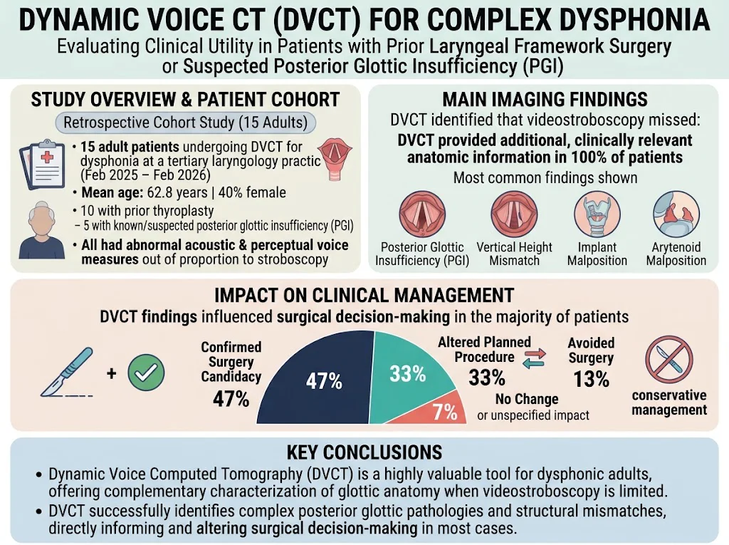
Dynamic Voice Computed Tomography (DVCT) significantly enhances the anatomical assessment of the glottis in adults presenting with dysphonia disproportionate to videostroboscopy findings. DVCT detects subtle structural abnormalities such as posterior glottic insufficiency, vertical height mismatch, and implant or arytenoid malposition. This imaging modality importantly influences clinical decision-making by confirming surgical candidacy, modifying planned procedures, or avoiding unnecessary surgery.
Study Background
Dysphonia, characterized by impaired voice quality, is a common complaint in otolaryngology, often attributed to vocal fold pathology. Videostroboscopy remains the gold standard for dynamic visualization of vocal fold vibration and mucosal wave activity. However, in certain complex cases— especially in adults with prior laryngeal framework surgery or suspected posterior glottic insufficiency (PGI)— videostroboscopy’s diagnostic capability can be limited. Anatomic alterations, supraglottic hyperfunction, or obscured posterior glottic view may impede adequate assessment of glottic function and structure. Posterior glottic insufficiency, marked by incomplete closure of the posterior vocal folds during phonation, may be subtle and underdiagnosed yet contributes substantially to dysphonia and breathiness. Precise anatomical delineation is crucial for tailoring appropriate surgical management. Dynamic Voice Computed Tomography (DVCT) is an emerging imaging technique that captures real-time anatomical motion of the larynx during phonatory tasks, potentially overcoming the limitations of videostroboscopy.
Study Design
This investigation was a retrospective cohort study conducted at a tertiary laryngology center between February 2025 and February 2026, involving 15 adult patients presenting with dysphonia disproportionate to videostroboscopic findings. The cohort had a mean age of 62.8 years and included 40% females. The population was enriched for patients with prior thyroplasty or those clinically suspected of PGI. Data collection encompassed demographics, detailed surgical histories, standardized acoustic and perceptual voice assessments, and videostroboscopic evaluation. All patients subsequently underwent DVCT to further characterize their glottic anatomy dynamically. The DVCT acquisition captured axial and volumetric images during phonation to assess glottic closure, vocal fold position, and any hardware or anatomic anomalies such as implant or arytenoid malpositions. The principal endpoint was to determine whether DVCT provided additional clinically pertinent anatomic information to guide therapeutic decisions beyond what videostroboscopy revealed.
Key Findings
All 15 patients displayed abnormal acoustic and perceptual voice measures that were disproportionate to stroboscopic findings, indicating the need for supplementary assessment. DVCT successfully yielded additional, clinically relevant anatomical data in every patient. The most frequently identified abnormalities were posterior glottic insufficiency, vertical height mismatch of the vocal folds, malposition of thyroplasty implants, and arytenoid cartilage malposition—findings that were either invisible or underestimated by videostroboscopy.
Clinically, DVCT findings had substantial impact on management: – Confirmation of surgical candidacy in 47% of patients, strengthening the indication for intervention. – Modification of planned surgical procedures in 33%, allowing more tailored and targeted approaches based on precise anatomical derangements. – Avoidance of surgery in 13%, preventing unwarranted intervention in patients whose anatomy suggested conservative management or alternative therapies.
These results underscore DVCT’s value as a complementary imaging tool, bridging key diagnostic gaps when conventional stroboscopy is ambiguous or insufficient, especially in post-surgical larynxes or cases of suspected posterior glottic insufficiency. Dynamic CT’s ability to visualize structural mobility, spatial relationships, and hardware position with high resolution enhances clinicians’ understanding of complex glottic pathophysiology.
Expert Commentary
Given that videostroboscopy remains limited by altered anatomy, edema, and supraglottic compensatory movements, DVCT represents a promising adjunct in voice medicine. By enabling objective evaluation of three-dimensional laryngeal architecture and movement, DVCT facilitates identification of subtle but functionally significant structural abnormalities underlying persistent dysphonia. Importantly, this can optimize patient selection for surgical intervention and tailor operative strategies, potentially resulting in improved voice outcomes.
While the study sample was modest and retrospective, its findings align with a growing body of literature endorsing advanced imaging modalities for complex laryngeal conditions. Future investigations should aim to define diagnostic criteria for DVCT, standardize protocols, and link imaging metrics with validated voice outcome measures prospectively. Additionally, balance between radiation exposure and diagnostic benefit must be carefully considered.
Conclusion
Dynamic Voice Computed Tomography is a valuable diagnostic addition for adult patients with dysphonia that is disproportionate to clinical and videostroboscopic findings, particularly in those with prior laryngeal framework surgery or suspected posterior glottic insufficiency. By providing enhanced visualization of glottic anatomy and motion, DVCT refines diagnostic accuracy and significantly informs surgical decision-making, improving individualized patient care. Further research should expand on its utility, optimize indications, and integrate DVCT into multimodal voice assessment algorithms.
Funding and Registration
The study was conducted at a tertiary laryngology practice; however, no specific funding or clinical trial registration details were reported.
References
1. Larson PJ, Schuman AD, Friedman AD, Dion GR, Burch MB, Howell RJ. Dynamic Voice Computed Tomography for Dysphonia Disproportionate to Clinical Findings. The Laryngoscope. 2026 Nov 2. PMID: 42972625.
2. Zeitels SM, Akst LM. Surgical Management of Posterior Glottic Stenosis: From Pathophysiology to Treatment. Otolaryngol Clin North Am. 2020;53(1):14-24.
3. Woo P, Ma EP, Ayad T. Videostroboscopy and its limitations in voice assessment: A review. J Voice. 2018;32(6):784-793.
4. Hirano M. Current concepts in treatment for voice disorders. Otolaryngol Clin North Am. 2016;49(6):1301-1315.
Specialty calculators
For clinical reference; use professional judgment.