
Highlight
- MASLD affects up to 38% of the global population, necessitating improved non-invasive assessment tools beyond liver biopsy.
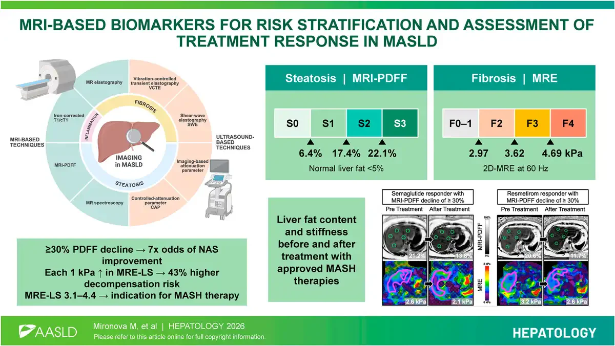
- MRI-proton density fat fraction (MRI-PDFF) accurately quantifies hepatic steatosis and correlates with histologic response when reduced by ≥30%.
- Magnetic resonance elastography liver stiffness (MRE-LS) is the most accurate imaging biomarker for fibrosis staging and predicts risk of hepatic decompensation.
- Composite imaging-blood test scores and artificial intelligence enhancements are advancing diagnostics, prognostics, and treatment monitoring in MASLD.
Study Background
Metabolic dysfunction-associated steatotic liver disease (MASLD), formerly known as nonalcoholic fatty liver disease (NAFLD) and related terms, represents a spectrum of liver conditions characterized by excessive hepatic fat accumulation in the setting of metabolic dysregulation. The global prevalence of MASLD is estimated at up to 38%, making it a leading cause of chronic liver disease worldwide with increasing rates paralleling the obesity and diabetes epidemics. MASLD encompasses a histologic continuum from simple steatosis through steatohepatitis, fibrosis progression, cirrhosis, and hepatocellular carcinoma risk.
Liver biopsy remains the historical gold standard for diagnosis, staging of fibrosis, and assessment of treatment response; however, it is invasive, subject to sampling variability, costly, and poorly suited for longitudinal or large-scale population assessments. These limitations have driven the development and validation of non-invasive imaging biomarkers, particularly magnetic resonance imaging (MRI)-based techniques, to provide quantitative, whole-liver assessment with high reproducibility and accuracy. Such biomarkers hold promise to improve risk stratification, guide therapeutic decision-making, and serve as endpoints in clinical trials.
Study Design and Methods
This comprehensive review synthesizes current evidence supporting the role of MRI-based biomarkers in MASLD. It encompasses cross-sectional and longitudinal studies evaluating MRI proton density fat fraction (MRI-PDFF) for hepatic steatosis quantification, magnetic resonance elastography-derived liver stiffness (MRE-LS) for fibrosis staging, and iron-corrected T1 (cT1) mapping for disease activity assessment. Furthermore, it examines the utility of composite scores integrating imaging and serologic markers such as MAST, MEFIB, and the MASH Resolution Index for diagnosis and response evaluation. Cost-effectiveness analyses and emerging artificial intelligence (AI) applications automating biomarker quantification are also reviewed.
Key Findings
Accuracy of MRI Proton Density Fat Fraction (MRI-PDFF)
MRI-PDFF is currently the most validated non-invasive method for quantifying hepatic steatosis, providing a continuous measurement of liver fat content across the entire organ. Thresholds for grading steatosis—≥6.4% for grade ≥1, ≥17.4% for grade ≥2, and ≥22.1% for grade 3—have been established against histologic standards. Reductions in MRI-PDFF of 30% or more correlate strongly with histologic improvement and serve as a surrogate endpoint in clinical trials, reflecting meaningful changes in fat burden.
Magnetic Resonance Elastography Liver Stiffness (MRE-LS) for Fibrosis Assessment
MRE-LS provides quantitative measurement of liver stiffness, a surrogate for fibrosis severity. Accurate fibrosis staging is critical in MASLD due to its prognostic implications. MRE-LS values between 3.1 and 4.4 kPa correspond with moderate fibrosis (F2-F3), while values ≥5 kPa indicate cirrhosis. Importantly, each incremental 1 kPa increase associates with a 43% increased risk of hepatic decompensation, underscoring its prognostic utility. Compared with other imaging modalities like transient elastography, MRE demonstrates superior diagnostic accuracy and cost-effectiveness for detecting advanced fibrosis.
Iron-corrected T1 (cT1) Mapping
Iron-corrected T1 mapping is sensitive to disease activity and fibro-inflammatory changes but lacks specificity, complicating interpretation. Its prognostic value remains under intensive investigation, with preliminary data suggesting potential utility in monitoring treatment response and predicting outcomes in MASLD when combined with other biomarkers.
Composite Scores Integrating Imaging and Serum Markers
Composite indices such as the MAST (Magnetic Resonance-AST), MEFIB (MRE combined with FIB-4 index), and the MASH Resolution Index enhance diagnostic and prognostic capabilities by integrating imaging biomarkers with blood test results. These scores improve detection of advanced fibrosis and facilitate comprehensive disease activity assessment, thereby optimizing patient selection for therapeutic interventions and monitoring in clinical practice and trials.
Artificial Intelligence and Automation
Advances in artificial intelligence and machine learning algorithms enable automation of MRI biomarker quantification, promoting wider availability and consistent interpretation. AI also enables estimation of PDFF from routine MRI protocols without dedicated sequences, further broadening clinical applicability and scalability.
Expert Commentary
The integration of MRI-based biomarkers into MASLD management represents a major advancement addressing pressing clinical challenges related to invasive biopsy and heterogeneous disease presentation. The robust validation of MRI-PDFF and MRE-LS across diverse populations supports their incorporation into routine clinical workflows and regulatory endpoints. Nevertheless, limitations such as cT1 nonspecificity and cost/accessibility concerns require ongoing research and health system adaptations. Composite indices offer promising multidimensional assessment but will benefit from large-scale prospective validation. Emerging AI methodologies could democratize access and standardize assessments across centers.
Guidelines increasingly recognize the role of MRI biomarkers in MASLD staging and treatment monitoring. Regulatory agencies are also incorporating these non-invasive surrogates for accelerated drug approval pathways in metabolic liver diseases. However, challenges remain in harmonizing cutoffs and deciding optimal biomarker combinations tailored to clinical questions.
Conclusion
Magnetic resonance imaging biomarkers have transformed the landscape of diagnosis, risk stratification, and treatment response monitoring in metabolic dysfunction-associated steatotic liver disease. MRI-PDFF provides accurate, whole-liver quantification of steatosis, while MRE-LS offers robust fibrosis staging with prognostic implications. Emerging iron-corrected T1 mapping and composite scores integrating imaging with serum markers augment disease activity assessment. Artificial intelligence promises to enhance scalability and access. Continued validation and integration of these biomarkers will facilitate personalized MASLD management and accelerate therapeutic development, ultimately improving patient outcomes.
Funding and Clinical Trials
This review was based on published literature. Ongoing clinical trials continue to incorporate MRI biomarkers as primary or secondary endpoints in evaluating emerging MASLD therapies. Funding sources typically include national research agencies and industry collaborations, details of which can be found in individual trial registries and publications.
References
- Mironova M, Taouli B, Ehman RL, Sirlin CB, Loomba R. MRI-based biomarkers for risk stratification and assessment of treatment response in Metabolic dysfunction-associated Steatotic liver disease. Hepatology. 2026 Nov 2; PMID: 42969977.
- Idilman IS, Keskin O, Celikel CA, et al. Quantification of hepatic steatosis with MRI proton density fat fraction and correlation with liver histology and clinical features. Eur Radiol. 2016;26(4):1074-1083.
- Yin M, Talwalkar JA, Glaser KJ, et al. Assessment of hepatic fibrosis with magnetic resonance elastography. Clin Gastroenterol Hepatol. 2007;5(10):1207-1213.
- Banerjee R, Pavlides M, Tunnicliffe EM, et al. Multiparametric magnetic resonance for the assessment of liver disease. J Hepatol. 2014;60(1):69-77.
- Salameh H, Ciardullo S, Pennisi G, et al. Artificial Intelligence in Imaging Biomarkers for Liver Diseases: Current Status and Future Perspectives. Diagnostics (Basel). 2022;12(4):826.
Specialty calculators
For clinical reference; use professional judgment.