Highlights
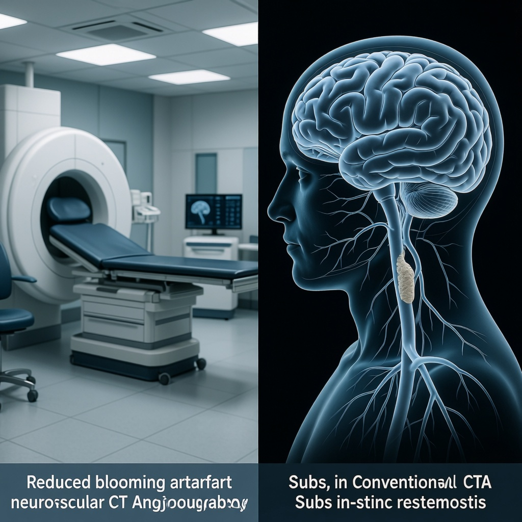
Ultra-high-resolution photon-counting detector CT angiography (UHR PCD-CTA) substantially improved image quality and reduced blooming artifacts compared with simulated conventional energy-integrating detector CTA.
In this prospective single-center cohort, UHR PCD-CTA detected neurovascular in-stent restenosis (ISR) with a sensitivity of 100.0%, specificity of 97.3%, and overall accuracy of 98.0% on a stent-based analysis.
Beyond restenosis detection, UHR PCD-CTA improved visualization of in-stent intimal hyperplasia and low-attenuation plaque in stents without restenosis, suggesting added value for surveillance of subclinical vessel wall changes.
The study supports photon-counting CTA as a promising noninvasive alternative for post-stent follow-up in neurovascular disease, although validation in larger multicenter cohorts remains necessary.
Clinical Background
Neurovascular stenting is used across a range of cerebrovascular conditions, including extracranial carotid stenosis, intracranial atherosclerotic disease, and selected aneurysm or arterial reconstruction procedures. A central challenge after stent implantation is surveillance for ISR, a process driven by neointimal hyperplasia, vessel wall remodeling, and sometimes progression of underlying atherosclerosis. Clinically important ISR may increase the risk of recurrent ischemic symptoms and can trigger repeat angiography or reintervention.
Digital subtraction angiography (DSA) remains the reference standard for evaluating in-stent luminal narrowing, but it is invasive, resource-intensive, and carries small yet meaningful risks, including stroke, contrast reactions, and access-site complications. For these reasons, noninvasive imaging strategies are highly desirable for longitudinal follow-up.
Conventional CTA has clear practical advantages, including broad availability and rapid acquisition, but assessment of metallic stents is limited by blooming artifacts and partial-volume effects. These issues can exaggerate the apparent thickness of stent struts and obscure the residual lumen, especially in small intracranial vessels where millimeter-level precision matters. Magnetic resonance angiography may also be degraded by susceptibility artifact, and duplex ultrasound has limited access to many intracranial segments. This leaves a persistent imaging gap in neurovascular ISR surveillance.
Photon-counting detector CT is an emerging technology that differs fundamentally from energy-integrating detector CT. Rather than converting x-rays into visible light before measuring total deposited energy, photon-counting systems register individual photons and their energy. This can improve spatial resolution, reduce electronic noise, and support spectral imaging. In the vascular setting, these properties raise the possibility of sharper delineation of the in-stent lumen and less artifact from metal. The study by Su and colleagues directly tests that clinical hypothesis in patients with prior neurovascular stent implantation.
Study Design
Design and setting
This was a prospective study conducted at The First Affiliated Hospital of Nanjing Medical University between July and November 2025. Consecutive patients with prior neurovascular stent implantation were enrolled, an approach that reduces some selection bias compared with retrospective case assembly.
Population
The final cohort included 43 participants with 51 neurovascular stents. The mean age was 63.1 years with a standard deviation of 9.5 years, and 8 participants were women. The analysis was performed at the stent level rather than purely at the patient level, which is appropriate when individual patients may contribute more than one treated vascular segment.
Imaging protocol and comparator
All participants underwent UHR PCD-CTA. To create a fair within-subject comparison against conventional CTA performance, the investigators reconstructed simulated energy-integrating detector CTA data sets from the same raw data. This design is methodologically appealing because it minimizes confounding from differences in timing, contrast enhancement, patient motion, and anatomy that would arise if two separate scans were compared.
Image assessment
Two blinded radiologists independently evaluated subjective image quality, diagnostic confidence for ISR, and the visibility of in-stent intimal hyperplasia and low-attenuation plaque in stents without restenosis. Although the abstract does not provide full scoring scales, the use of blinded readers is an important strength when the main outcomes include image-based interpretation.
Reference standard and endpoints
The principal clinical endpoint was diagnostic performance for ISR detection on a stent-based level. DSA served as the reference standard when available. DSA was available for 26 of 51 stents, including all stents with ISR. This means sensitivity estimates for detecting diseased stents were anchored to invasive confirmation, but not every non-ISR case underwent DSA verification.
Key Findings
Image quality and artifact reduction
Compared with simulated conventional CTA, UHR PCD-CTA produced significantly higher subjective image quality, with a reported P value of 0.004. It also yielded significantly greater diagnostic confidence for identifying ISR, with P<0.001. Most notably, blooming artifacts were markedly reduced, also with P<0.001.
These findings are clinically important because the major barrier to CT-based stent evaluation has not been merely image aesthetics, but the inability to distinguish artifact from true luminal compromise. A technically better image only matters if it supports more reliable diagnostic decisions. The parallel gain in reader confidence suggests that the improved appearance translated into more actionable interpretation.
Diagnostic performance for ISR
Among 51 stents, ISR was present in 14 and absent in 37. UHR PCD-CTA achieved a sensitivity of 100.0%, specificity of 97.3%, and overall accuracy of 98.0% for ISR detection on a stent-based basis. In practical terms, the technique identified all stents with restenosis in this cohort and generated very few false-positive interpretations among stents without ISR.
Given the clinical consequences of missed ISR, sensitivity is particularly important. A sensitivity of 100% is encouraging, although it must be interpreted cautiously because it is based on 14 positive stents. With such a modest event count, the confidence interval around sensitivity would likely be relatively wide even if not reported in the abstract. Still, the absence of false negatives in this early experience is notable.
The specificity of 97.3% is also impressive. Conventional CTA often struggles with false positives because metal-related blooming narrows the apparent lumen. Reducing those false positives could lessen unnecessary invasive angiography and reassure clinicians when routine surveillance imaging appears normal.
Visualization of in-stent vessel wall pathology
In stents without restenosis, UHR PCD-CTA significantly improved visualization of in-stent intimal hyperplasia and low-attenuation plaque compared with simulated energy-integrating detector CTA, with P<0.001. This is more than a secondary technical observation. It suggests that photon-counting CTA may not only classify stents as restenotic or patent, but may also characterize early structural changes within the stented segment before a hemodynamically significant stenosis develops.
If reproduced, this capability could reshape surveillance strategies. Rather than a binary search for advanced restenosis, imaging follow-up might evolve toward earlier detection of biologic activity within the stent, potentially informing antiplatelet management, lipid-lowering intensification, or closer interval follow-up in selected patients. That said, the prognostic significance of these imaging findings remains uncertain and would need dedicated longitudinal study.
Why Photon-Counting CT May Matter Mechanistically
The performance gains reported here are biologically and physically plausible. Photon-counting CT offers smaller detector elements and improved intrinsic spatial resolution, helping separate thin metal struts from contrast-opacified lumen. It can also reduce electronic noise and improve contrast-to-noise characteristics. Together, these features are especially useful in neurovascular applications, where arteries are smaller than coronary or peripheral vessels and where a submillimeter loss of clarity can obscure clinically relevant narrowing.
The technology may also open future opportunities in spectral analysis, material decomposition, and virtual monoenergetic imaging, which could further optimize vessel visualization and metal artifact handling. Although the current study focused primarily on UHR reconstruction and comparison with simulated conventional CTA, the platform’s broader spectral capabilities may be relevant in future neurovascular protocols.
Clinical Interpretation
This study addresses a meaningful unmet need. In current practice, clinicians often face a trade-off between imperfect noninvasive surveillance and invasive angiographic certainty. For intracranial or extracranial neurovascular stents, especially in patients requiring repeated follow-up, a noninvasive test with high sensitivity and high specificity would be valuable.
The results suggest that UHR PCD-CTA could move CTA closer to that goal. If these findings are confirmed, several clinical use cases are conceivable. First, it could become a preferred first-line test for routine post-stent surveillance in stable patients. Second, it might reduce the number of diagnostic DSAs performed solely to clarify equivocal CTA findings. Third, it may help identify subtle in-stent tissue changes that warrant intensified risk-factor modification even before restenosis becomes severe.
For neurologists, neurointerventionalists, and vascular radiologists, the most compelling aspect is not simply the high accuracy number, but that the study tested a real-world clinical problem where conventional CTA often disappoints. The use of raw-data-derived simulated conventional CTA as the comparator strengthens the inference that the observed gains are attributable to detector technology and ultra-high-resolution reconstruction rather than scan timing or patient heterogeneity.
Limitations and Remaining Questions
Despite its promise, this is still an early study and should be interpreted as such. The sample size was modest: 43 patients and 51 stents, with only 14 restenotic stents. Performance estimates from small cohorts can appear excellent yet shift materially in larger populations.
The study was performed at a single center, which may limit generalizability. Scanner-specific protocols, reconstruction techniques, and reader experience may influence results. Broader multicenter evaluation will be necessary to determine whether similar performance can be achieved across institutions and in more heterogeneous patient groups.
Another important caveat is the reference standard structure. DSA was available for 26 stents, including all stents with ISR, but not all non-ISR stents were verified invasively. This introduces the possibility of partial verification bias. While understandable from an ethical and practical standpoint, because routine invasive angiography in clearly normal cases is hard to justify, it means specificity estimates should be interpreted with some caution.
The abstract does not report inter-reader agreement, radiation dose metrics, contrast dose details, confidence intervals, or ISR severity thresholds. These are not trivial omissions for implementation. Clinicians will want to know whether the improved image quality comes with comparable or lower dose than conventional CTA, whether performance varies by stent type or diameter, and how reproducible the interpretations are between readers.
It is also unclear how photon-counting CTA compares with high-resolution flat-panel CT, MRA techniques, duplex ultrasound for extracranial segments, or combined multimodality follow-up strategies. Cost, scanner availability, workflow integration, and reimbursement will influence adoption as much as diagnostic performance.
Relation to the Existing Literature
The current findings fit with a broader radiology literature showing that photon-counting CT can improve visualization of fine anatomic detail and reduce limitations associated with small structures and high-contrast interfaces. Prior work in coronary imaging, temporal bone imaging, and lung imaging has demonstrated the technical advantages of photon-counting detectors, including enhanced spatial resolution and spectral capability. Neurovascular stent surveillance is a logical extension of these strengths.
Within cerebrovascular practice, guideline-level evidence for optimal noninvasive ISR surveillance remains limited, partly because imaging technologies have had inconsistent performance in the presence of metallic implants. Therefore, studies such as this one are important not only as technology demonstrations but also as potential foundations for future care pathways.
Practical Takeaways for Clinicians
For now, UHR PCD-CTA should be viewed as a highly promising advanced imaging option rather than an established standard of care. In centers with access to photon-counting CT, it may already offer a useful adjunct for difficult post-stent cases, especially when conventional CTA is equivocal and DSA is being considered primarily for clarification.
When interpreting these results in practice, clinicians should keep three points in mind. First, the technique appears particularly strong at minimizing false reassurance risk, since all ISR cases in this cohort were detected. Second, improved visualization of intimal hyperplasia may generate new findings whose management implications are not yet fully defined. Third, local expertise in acquisition and reconstruction will likely matter; a sophisticated scanner does not eliminate the need for optimized protocols and experienced readers.
Conclusion
The study by Su CQ, Zhou C, Wu PF, Li Q, Zhang T, Chen JL, Xu XQ, Shi HB, Liu S, Wu FY, and Lu SS provides compelling early evidence that ultra-high-resolution photon-counting CTA can substantially improve noninvasive evaluation of neurovascular stents. Compared with simulated conventional CTA, it improved image quality, reduced blooming artifact, increased diagnostic confidence, and achieved excellent stent-level performance for ISR detection.
If validated in larger, multicenter studies with comprehensive reference standards and workflow data, UHR PCD-CTA could become an important tool for post-stent surveillance in neurovascular disease. Its potential to depict not only overt restenosis but also earlier in-stent tissue changes makes it especially interesting. At present, the technology appears poised to narrow one of the most persistent diagnostic gaps in cerebrovascular imaging: accurate, noninvasive assessment of the stented lumen.
Funding and Trial Registration
The abstract provided does not report funding information or a ClinicalTrials.gov registration number. Readers should consult the full published article for these details.
References
1. Su CQ, Zhou C, Wu PF, Li Q, Zhang T, Chen JL, Xu XQ, Shi HB, Liu S, Wu FY, Lu SS. Ultra-High-Resolution Photon-Counting CT Angiography for Noninvasive Detection of Neurovascular In-Stent Restenosis. Stroke. 2026 Jun 2. PMID: 42227106.
2. Willemink MJ, Persson M, Pourmorteza A, Pelc NJ, Fleischmann D. Photon-counting CT: technical principles and clinical prospects. Radiology. 2018;289(2):293-312.
3. Flohr T, Petersilka M, Henning A, Ulzheimer S, Ferda J, Schmidt B. Photon-counting CT review. Phys Med. 2020;79:126-136.
4. Saba L, Raz E, Grassi R, Wintermark M. Imaging of the carotid artery vulnerable plaque. Cardiovasc Intervent Radiol. 2014;37(3):572-585.
5. Marks MP, Marcellus ML, Do HM, Schraedley-Desmond PK, Steinberg GK, Tong DC, Albers GW. Intracranial angioplasty without stenting for symptomatic atherosclerotic stenosis: long-term follow-up. AJNR Am J Neuroradiol. 2005;26(3):525-530.