![Persistent Fibroblast Activation Predicts Heart Failure Progression: [68Ga]FAPI-46 PET/MRI Reveals Distinct Patterns in Ischemic and Nonischemic Cardiomyopathy](https://news.medxy.ai/wp-content/uploads/2026/04/d43c8ff0-6629-4944-bc15-78efd79810d7.jpg)
Background: The Fibrosis Paradox in Heart Failure
Heart failure remains a global epidemic, affecting over 64 million people worldwide and carrying a five-year mortality rate that rivals many malignancies. While significant advances have been made in pharmacological and device-based therapies, a substantial proportion of patients continue to experience progressive deterioration despite optimal medical management. This clinical reality has prompted researchers to look beyond conventional hemodynamic parameters and explore the underlying biological drivers of adverse cardiac remodeling.
At the cellular level, myocardial fibrosis represents one of the most critical pathobiological processes contributing to heart failure progression. Activated cardiac fibroblasts play a central role in this process, depositing extracellular matrix proteins and contributing to ventricular stiffness, diastolic dysfunction, and arrhythmogenesis. However, the extent and pattern of fibroblast activation vary considerably between patients, particularly when comparing ischemic cardiomyopathy (ICM) resulting from myocardial infarction to nonischemic cardiomyopathy (NICM) with diverse etiologies such as inflammatory, toxic, or idiopathic causes.
Noninvasive assessment of fibroblast activation has historically presented a significant diagnostic challenge. While late gadolinium enhancement (LGE) cardiac magnetic resonance can identify established scar tissue, it does not directly visualize active fibrogenic processes occurring in both scarred and non-scarred myocardial regions. This limitation has hindered our ability to phenotype patients based on their underlying biological activity and predict which individuals might benefit from emerging antifibrotic therapies.
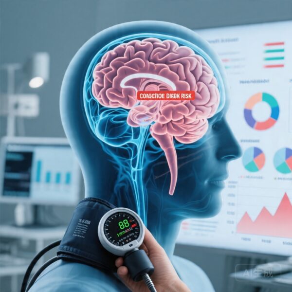
The recent development of gallium 68-labeled fibroblast activation protein inhibitor ([68Ga]FAPI-46) positron emission tomography represents a paradigm shift in cardiac molecular imaging. Fibroblast activation protein (FAP) is a cell surface enzyme highly expressed by activated fibroblasts in various pathological conditions, making it an attractive target for imaging. The study by Joshi and colleagues, published in JAMA Cardiology, represents the first comprehensive application of this technology to evaluate myocardial fibroblast activation across the spectrum of heart failure with reduced ejection fraction (HFrEF).
Study Design: A Prospective Multicenter Investigation
This prospective case-control study enrolled 81 participants from multiple cardiac centers, with data analysis conducted between January 2024 and January 2025. The study population was carefully stratified into four distinct groups to enable meaningful comparisons across the heart failure spectrum.
The primary cohort consisted of 42 patients with HFrEF, further divided into two etiologic subgroups: 21 patients with heart failure due to ischemic cardiomyopathy (all with prior myocardial infarction) and 21 patients with nonischemic cardiomyopathy etiologies. These patients had a mean left ventricular ejection fraction of 41% with a standard deviation of 9%, representing moderate to severe systolic dysfunction. A secondary comparison group included 20 patients with prior myocardial infarction but preserved left ventricular systolic function, allowing researchers to isolate the effects of infarction from those of overt heart failure. Finally, 19 age-matched healthy volunteers served as controls to establish baseline reference values.
All participants underwent comprehensive [68Ga]FAPI-46 PET/MRI imaging, which combined the metabolic signal of fibroblast activation with the anatomical and tissue characterization capabilities of magnetic resonance imaging. A subset of heart failure patients underwent repeat imaging after more than six months to evaluate longitudinal changes in fibroblast activation and their relationship to clinical outcomes.
The primary endpoint was myocardial fibroblast activation quantified using maximum standardized uptake values (SUVmax) of [68Ga]FAPI-46. Secondary analyses explored the spatial distribution of uptake, correlations with left ventricular remodeling parameters, and associations between baseline activation levels and subsequent changes in ejection fraction with optimal medical therapy.
Key Findings: Distinct Patterns of Fibroblast Activation Emerge
The results of this investigation revealed striking differences in myocardial fibroblast activation that closely mirrored the underlying cardiac pathology. Most notably, healthy volunteers demonstrated essentially no myocardial [68Ga]FAPI-46 uptake, establishing a clear biological boundary between physiological aging and pathological fibrogenesis.
In contrast, all patients with heart failure exhibited significantly elevated myocardial [68Ga]FAPI-46 uptake compared to healthy controls. The mean SUVmax in the heart failure cohort was 2.7 with a standard deviation of 1.5, compared to 1.5 with a standard deviation of 0.3 in healthy volunteers, achieving high statistical significance (P < .001). This finding confirms that persistent fibroblast activation characterizes the failing human heart and is not merely a residual finding from past injury events.
Perhaps the most clinically relevant observation concerned the distinct spatial patterns observed between ischemic and nonischemic etiologies. Patients with ischemic cardiomyopathy demonstrated the highest myocardial uptake, with [68Ga]FAPI-46 signal localizing precisely to regions of established myocardial infarction. The mean SUVmax in this subgroup reached 3.2 with a standard deviation of 1.1, significantly higher than all other groups. This pattern is consistent with chronic wound healing processes ongoing within scar tissue, where fibroblasts continue to remodel and maintain the fibrotic scar years after the initial ischemic event.
Patients with nonischemic cardiomyopathy exhibited a markedly different pattern of fibroblast activation. Rather than focal uptake in scar regions, these patients demonstrated diffuse, lower-intensity uptake distributed throughout the myocardium. The mean SUVmax of 2.3 with a standard deviation of 0.5 was significantly lower than the ischemic group, but notably, the highest signal was consistently observed in the basal septum regardless of whether late gadolinium enhancement was present. This observation suggests that nonischemic fibrotic processes preferentially affect specific myocardial regions and can occur independently of visible scar formation on conventional imaging.
An intriguing finding emerged from the comparison between heart failure patients with ischemic etiology and patients with prior myocardial infarction who had not developed heart failure. Despite having no major difference in overall infarct size, ischemic cardiomyopathy patients with heart failure demonstrated significantly higher [68Ga]FAPI-46 uptake than those without heart failure (mean SUVmax 3.2 versus 2.5, P = .03). This discordance between structural injury extent and biological activity suggests that additional factors beyond scar size determine which patients progress to overt heart failure, potentially including the intensity and persistence of fibrogenic signaling.
The longitudinal analysis provided perhaps the most compelling evidence for the clinical significance of fibroblast activation imaging. Among heart failure patients who underwent repeat imaging, higher baseline [68Ga]FAPI-46 uptake was strongly associated with less improvement in left ventricular ejection fraction despite optimal medical therapy over time (r = -0.52, P = .02). This correlation indicates that the magnitude of ongoing fibrogenic activity at baseline can predict which patients will respond poorly to conventional treatment, offering a potential avenue for risk stratification and personalized therapeutic targeting.
Expert Commentary: Implications for Heart Failure Management
These findings carry substantial implications for the understanding and management of heart failure. The demonstration of persistent fibroblast activation in heart failure patients, regardless of etiology, provides biological rationale for the progressive nature of the disease despite apparent hemodynamic optimization. Activated fibroblasts not only deposit collagen but also secrete cytokines, growth factors, and matrix metalloproteinases that collectively influence cardiac geometry, coronary microvascular function, and electrical stability.
The distinct spatial patterns observed between ischemic and nonischemic cardiomyopathies suggest fundamentally different fibrogenic mechanisms may be operating. In ischemic cardiomyopathy, the pattern reflects chronic inflammation and wound remodeling within established scar tissue. In nonischemic cardiomyopathy, the diffuse distribution with basal septal predominance may relate to different pathophysiological drivers such as microvascular dysfunction, inflammatory activation, or genetic predisposition affecting specific myocardial layers.
The observation that baseline fibroblast activation predicts treatment response represents a potential breakthrough in phenotyping heart failure patients. Currently, clinicians rely primarily on ejection fraction, functional class, and biomarker levels to guide prognosis and therapy selection. The addition of FAP-targeted imaging could enable identification of patients with predominant fibrotic phenotypes who might derive particular benefit from emerging antifibrotic agents currently in development, including transforming growth factor-beta pathway inhibitors, pirfenidone, and nintedanib.
Several limitations warrant consideration when interpreting these findings. The sample size, while adequate for demonstrating proof-of-concept, limits subgroup analyses and prevents robust evaluation of rare adverse events. The single-timepoint nature of the primary imaging protocol means that the temporal dynamics of fibroblast activation throughout heart failure progression remain incompletely characterized. Additionally, the study population was predominantly male and older, raising questions about generalizability to younger patients, women, and populations with different genetic backgrounds or comorbidities.
Conclusion: A New Window into Cardiac Fibrosis
This landmark study establishes [68Ga]FAPI-46 PET/MRI as a powerful tool for noninvasive assessment of myocardial fibroblast activation in heart failure. The demonstration of persistent activation with distinct spatial patterns according to cardiomyopathy etiology provides mechanistic insights into disease heterogeneity and progression. Perhaps most importantly, the correlation between baseline activation and subsequent treatment response offers a new paradigm for risk stratification and personalized therapeutic decision-making.
As antifibrotic therapies advance through clinical development, the ability to image and quantify active fibrogenesis becomes increasingly critical for patient selection, dose optimization, and treatment monitoring. Future studies should validate these findings in larger, more diverse populations and explore whether serial imaging can guide therapeutic interventions. The path toward targeted antifibrotic therapy for heart failure now has a molecular imaging companion that may ultimately improve outcomes for millions of patients worldwide.
Funding and Clinical Trials
This research was conducted as a prospective observational study registered with clinicaltrials.gov. The study received support from multiple academic and institutional sources including the University of Edinburgh, NHS Research Scotland, and the British Heart Foundation. Specific grant numbers and institutional affiliations are documented in the original publication.
References
Joshi SS, Barton AK, Loganath K, et al. Myocardial Fibroblast Activation in Ischemic and Nonischemic Cardiomyopathy. JAMA Cardiology. 2026. PMID: 41984469.
Aghajanian H, Kimura T, Rurik JG, et al. Targeting cardiac fibrosis with engineered T cells. Nature. 2019.
Dobaczewski M, de Haan JJ, Frangogiannis NG. The extracellular matrix modulates fibroblast phenotype and function in the infarcted myocardium. J Mol Cell Cardiol. 2016.
Gao E, Lei YH, Shang X, et al. Accurate and efficient approach to quantify myocardial fibrosis with extracellular volume fraction from routine clinical LGE. JACC Cardiovasc Imaging. 2018.