Highlights
Uterine leiomyomas remain common through midlife and do not predictably disappear after menopause. Longitudinal data suggest heterogeneous behavior: some fibroids shrink, some remain stable, and a subset continue to enlarge.
In perimenopausal and postmenopausal patients, abnormal uterine bleeding and pelvic pain should not be attributed to fibroids by default. Endometrial pathology and other gynecologic or nongynecologic causes require systematic exclusion.
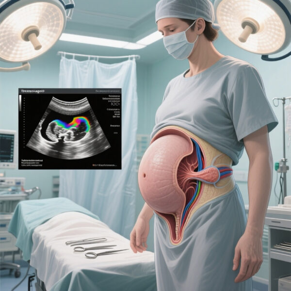
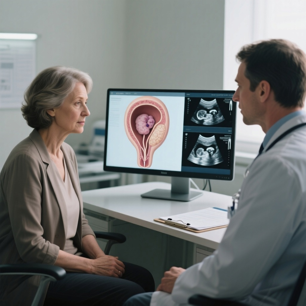
Transvaginal ultrasonography remains the first-line imaging test, while magnetic resonance imaging is useful when sonographic findings are indeterminate, anatomy is complex, or concern for malignancy is higher.
No individual imaging feature can reliably distinguish leiomyoma from leiomyosarcoma. Although unsuspected leiomyosarcoma at surgery is uncommon, risk rises with age, making clinical context and careful preoperative counseling essential.
Background and Clinical Burden
Uterine leiomyomas are the most common solid pelvic tumors in women and remain a major cause of abnormal uterine bleeding, pelvic pressure, pain, bulk symptoms, infertility, and reduced quality of life. Their cumulative burden peaks during the reproductive years but often extends into the menopausal transition, when clinicians and patients may mistakenly expect spontaneous regression. That expectation is partly rooted in the hormone responsiveness of leiomyomas, which are influenced by ovarian steroids. Yet real-world clinical behavior is more variable than the classic teaching suggests.
The review by Encalada-Soto, Hochberg, and Laughlin-Tommaso addresses this clinically important gap. It focuses on how leiomyomas behave in perimenopausal and postmenopausal patients and how clinicians should approach evaluation when symptoms arise in an age group where endometrial neoplasia, adnexal pathology, and uterine sarcoma become more relevant diagnostic considerations.
The disease burden is not evenly distributed. Racial disparities remain a defining epidemiologic feature of leiomyoma care. Black women tend to experience earlier onset, larger uterine volume, more numerous tumors, greater symptom burden, and higher rates of intervention. These disparities persist into midlife and shape both baseline risk and treatment trajectories during the menopausal transition.
Article Type and Evidence Base
This publication is a structured, nonsystematic review of contemporary literature. According to the abstract, the authors searched PubMed and Embase and supplemented the search with guidelines and reference review. As such, the article is best understood as a clinically oriented synthesis rather than a formal systematic review or meta-analysis.
That distinction matters. A structured nonsystematic review is highly useful for practice, especially when it integrates imaging, symptom assessment, and age-specific management. However, it is not designed to provide pooled effect estimates, risk-of-bias grading, or exhaustive evidence capture. The value of the paper lies in its pragmatic interpretation of heterogeneous data and its focus on diagnostic reasoning in a population where symptom attribution can be misleading.
Proposed Clinical Structure for Evaluating Leiomyomas Across Menopause
The topic lends itself to a practical framework built around five questions: What is the expected biology of fibroids near menopause? Which symptoms should trigger further workup? What is the role of imaging? How should clinicians think about malignancy risk? And when is treatment necessary?
This structure is more clinically useful than a purely disease-centered overview because menopausal status changes both the differential diagnosis and the threshold for intervention.
Key Findings
Leiomyoma behavior after menopause is variable, not uniform
The central message of the review is that menopause does not guarantee fibroid resolution. While estrogen and progesterone decline generally favor regression, longitudinal observations show mixed trajectories. Some leiomyomas shrink modestly, others remain essentially unchanged, and some continue to grow despite ovarian senescence. This variability likely reflects differences in tumor biology, local steroid metabolism, receptor expression, vascularity, degeneration patterns, and exogenous hormonal exposure.
For frontline clinicians, the practical implication is straightforward: persistence of a fibroid after menopause is not, by itself, evidence of malignancy, but continued growth or new symptoms should not be dismissed as biologically implausible. Menopause changes probabilities; it does not eliminate heterogeneity.
Symptoms in perimenopause and postmenopause require a broader differential diagnosis
Abnormal uterine bleeding in the menopausal transition is common and often multifactorial. In perimenopause, anovulation and structural pathology frequently coexist. In postmenopause, any bleeding episode warrants evaluation because endometrial hyperplasia and endometrial carcinoma become more important considerations. Leiomyomas may still contribute to bleeding, but they should not be presumed to be the cause without appropriate assessment.
Pelvic pain and pressure similarly require careful interpretation. Bulk-related discomfort from fibroids can persist into later life, but adnexal masses, pelvic floor disorders, urinary tract pathology, gastrointestinal conditions, and degenerative or musculoskeletal pain may present similarly. The review appropriately emphasizes systematic evaluation over reflex attribution.
Transvaginal ultrasonography remains foundational
Transvaginal ultrasonography is the first-line imaging modality for suspected uterine leiomyomas across the menopausal transition. It is widely available, relatively inexpensive, and well suited to define uterine size, myometrial masses, distortion of the endometrial cavity, and competing pelvic pathology. A structured ultrasound assessment should document the number, size, location, and echotexture of lesions, as well as endometrial thickness when clinically relevant.
For postmenopausal bleeding, ultrasound has a second role beyond fibroid assessment: evaluation of the endometrium. Even when fibroids are present, clinicians must still decide whether endometrial sampling is indicated based on bleeding pattern, endometrial appearance, risk factors, and imaging limitations.
MRI is an adjunct, not a replacement for first-line assessment
Magnetic resonance imaging is particularly helpful when sonographic findings are limited by uterine size, numerous masses, atypical degeneration, poor acoustic windows, or uncertainty about lesion origin. MRI can better define tissue planes, characterize degeneration, assess the endometrial cavity, and clarify whether an apparent uterine mass may instead be adnexal or extrauterine.
Importantly, the review does not overstate MRI’s diagnostic certainty. MRI improves anatomic and tissue characterization, but even high-quality MRI cannot definitively exclude leiomyosarcoma. This is a critical and often misunderstood point in clinical practice.
No single imaging feature rules in or rules out leiomyosarcoma
The distinction between benign leiomyoma and leiomyosarcoma remains one of the most difficult problems in gynecologic imaging. Suspicious features such as rapid enlargement, heterogeneous signal, irregular margins, central necrosis, or abnormal diffusion characteristics may heighten concern, but none is pathognomonic. Benign leiomyomas with degeneration can look worrisome, while sarcomas can overlap with benign tumors on imaging.
The review reinforces a balanced message: the prevalence of unsuspected leiomyosarcoma at surgery is low, but age increases risk. Therefore, preoperative evaluation should integrate age, symptom pattern, growth history, imaging findings, menopausal status, and procedural consequences, especially when considering tissue extraction methods or minimally invasive approaches.
Management should be individualized and symptom driven
The article supports an individualized approach anchored in symptoms, diagnostic certainty, comorbidities, and patient preferences. Expectant management is appropriate for asymptomatic patients, including many postmenopausal women with stable fibroids identified incidentally. In contrast, intervention becomes reasonable when symptoms are persistent or severe, when quality of life is impaired, when anemia develops, or when uncertainty about diagnosis remains unresolved.
This symptom-driven framework is especially relevant in later reproductive life, when competing health priorities, medical comorbidities, anticoagulant use, obesity, and patient treatment goals may make conservative management more or less attractive. The decision is not simply whether a fibroid exists, but whether it is causing clinically meaningful disease and whether the anticipated benefit of treatment outweighs procedural and anesthetic risk.
Clinical Interpretation
Why the old assumption of universal postmenopausal regression is no longer sufficient
Traditional teaching linked leiomyoma behavior closely to ovarian hormone withdrawal. That model remains directionally true but clinically incomplete. Fibroids are biologically heterogeneous tumors with variable extracellular matrix composition, aromatase activity, receptor signaling, and blood supply. In addition, the menopausal transition itself is hormonally unstable rather than abrupt. Intermittent ovarian activity, obesity-related peripheral estrogen production, and exogenous hormone exposure may all contribute to continued symptomatology or lesion persistence.
For clinicians, the key lesson is to avoid two opposite errors: assuming all fibroids should shrink and therefore overinvestigating every persistent lesion, or assuming a known fibroid explains all symptoms and therefore missing endometrial disease or another pelvic disorder.
How to approach postmenopausal bleeding when fibroids are present
The presence of fibroids can complicate but should not derail the standard evaluation of postmenopausal bleeding. If the endometrium cannot be adequately visualized on ultrasound because of cavity distortion or shadowing, additional imaging or endometrial sampling may be necessary. In women with persistent or recurrent bleeding, reassuring imaging alone may not be enough. The review’s practical message aligns with guideline-based care: symptom-led reassessment remains essential.
When growth becomes concerning
Growth alone is not a definitive marker of sarcoma, but growth in a postmenopausal patient should prompt renewed diagnostic scrutiny. The degree, tempo, and context matter. A slowly enlarging mass in a woman using menopausal hormone therapy may carry a different pretest probability than a rapidly enlarging solitary uterine mass in a patient with new pain, constitutional symptoms, or imaging heterogeneity. This is an area where experienced clinical judgment remains indispensable because evidence has not yielded a single reliable algorithm.
Management Considerations Across the Menopausal Transition
Expectant management
For asymptomatic or minimally symptomatic patients, observation is often appropriate. This approach is particularly reasonable when imaging is reassuring, symptoms are stable, and the patient prefers to avoid intervention. Follow-up should be individualized rather than automatic, with reassessment driven by new bleeding, pressure symptoms, interval enlargement, or evolving patient preferences.
Medical therapy
Medical treatment in perimenopause may be used to reduce bleeding and improve quality of life while patients approach natural menopause. Options may include hormonal therapies or nonhormonal bleeding management depending on the patient’s risk profile and need for contraception. In postmenopause, the role of medical therapy is narrower and often shaped by whether the patient is using menopausal hormone therapy. Persistent symptoms in a postmenopausal patient generally warrant a renewed diagnostic review before simply escalating symptom treatment.
Procedural and surgical management
Procedural treatment, including hysterectomy or selected uterus-sparing options, remains appropriate for refractory symptoms or when diagnostic ambiguity persists. In patients near or after menopause, the threshold for definitive surgery may be influenced by completed childbearing, coexistence of adenomyosis or prolapse, recurrent bleeding, severe bulk symptoms, or concern about occult malignancy. However, the low prevalence of unsuspected leiomyosarcoma means that decisions should remain individualized rather than fear-driven.
Preoperative counseling should explicitly address uncertainty. Patients should understand that imaging cannot fully exclude sarcoma, that risk rises with age but remains low, and that the surgical route and tissue extraction method may be influenced by this background risk.
Strengths and Limitations of the Review
A major strength of the article is its clinical relevance. It moves beyond the simplistic question of whether fibroids shrink after menopause and instead addresses the real diagnostic and management problems clinicians face: bleeding, pain, indeterminate imaging, and malignancy concern. The emphasis on structured ultrasound, selective MRI use, and symptom-driven care is practical and consistent with modern gynecologic practice.
The limitations follow from the article type. As a nonsystematic review, it may not capture the full breadth of literature or quantify risk with the rigor of a formal evidence synthesis. The abstract does not report pooled rates, effect sizes, or evidence grading, so readers seeking precise estimates of postmenopausal growth frequency or age-stratified sarcoma prevalence will need to consult the primary literature and guideline documents directly.
Implications for Practice
This review supports several immediate practice points. First, persistent fibroids after menopause are not unusual. Second, new bleeding or pain in midlife and later life should trigger standard evaluation rather than symptom attribution alone. Third, transvaginal ultrasonography remains the core imaging test, with MRI reserved for clarification. Fourth, no imaging pattern can conclusively separate leiomyoma from leiomyosarcoma. Finally, management should be based on symptoms, risk context, and patient goals rather than assumptions about what menopause should do to fibroids.
These points are especially relevant in primary care, gynecology, radiology, and perioperative counseling. They also matter for health equity. Because leiomyoma burden and treatment exposure are not evenly distributed across populations, a nuanced approach to menopause-related symptom evaluation may help reduce both overtreatment and missed diagnoses.
Conclusion
Uterine leiomyomas across the menopausal transition demand a more nuanced framework than the longstanding expectation of universal regression. Contemporary evidence, as synthesized by Encalada-Soto and colleagues, indicates that fibroid behavior is heterogeneous, symptom evaluation must remain systematic, and imaging should be used thoughtfully rather than as a shortcut to certainty. The practical message is clear: asymptomatic patients can often be observed, but bleeding, pain, enlargement, or diagnostic ambiguity warrant careful reassessment. In this setting, individualized care is not merely preferable; it is the clinically responsible standard.
Funding and Trial Registration
No ClinicalTrials.gov registration applies to this structured review article. Funding information was not provided in the abstract available via PubMed and should be confirmed from the full publication.
References
1. Encalada-Soto D, Hochberg LS, Laughlin-Tommaso S. Uterine Leiomyomas Across the Menopausal Transition. Obstetrics and gynecology. 2026-06-04. PMID: 42241706.
2. Stewart EA, Laughlin-Tommaso SK, Catherino WH, Lalitkumar S, Gupta D, Vollenhoven B. Uterine fibroids. N Engl J Med. 2016;374(17):1646-1655.
3. Bulun SE. Uterine fibroids. N Engl J Med. 2013;369(14):1344-1355.
4. Practice Bulletin No. 228: Management of Symptomatic Uterine Leiomyomas. Obstet Gynecol. 2021;137(6):e100-e115.
5. Laughlin-Tommaso SK, Stewart EA. Moving toward individualized medicine for uterine leiomyomas. Obstet Gynecol Clin North Am. 2015;42(1):115-126.