Section Structure
Proposed article framework
1. Clinical context and unmet need in indeterminate pulmonary nodules. 2. Why geography matters: cancer risk versus endemic fungal disease. 3. Study design, cohorts, biomarkers, and statistical approach. 4. Main diagnostic findings and risk reclassification. 5. Clinical interpretation and potential workflow implications. 6. Strengths, limitations, and questions for validation. 7. Bottom line for practice and research. 8. Funding, registration, and references.
Highlights
Adding serum cancer proteins and Histoplasma antibodies to a standard clinical prediction tool improved diagnostic discrimination for indeterminate pulmonary nodules in two geographically distinct cohorts.
The full combined model achieved AUCs of 0.80 in the Ohio River Valley cohort and 0.79 in the Mountain West cohort, outperforming the Mayo model alone in both settings.
The approach appears particularly relevant for intermediate-risk nodules, where decision-making is often most difficult and unnecessary invasive procedures are common.
By incorporating fungal serology, the model addresses a major real-world confounder in endemic regions where benign granulomatous disease can mimic lung cancer.
Background
Indeterminate pulmonary nodules are a common and increasingly consequential problem in clinical practice. They are detected during lung cancer screening, incidental chest imaging, and workups for respiratory symptoms. Although many nodules are benign, a clinically important proportion represent early lung cancer, making accurate discrimination essential. The consequences of error run in both directions: underestimation can delay cancer diagnosis, while overestimation can lead to unnecessary biopsies, surgery, anxiety, and cost.
Current evaluation often begins with a validated clinical prediction model, such as the Mayo Clinic model, which uses variables like age, smoking history, prior extrathoracic cancer, nodule size, spiculation, and upper-lobe location. These tools are useful but imperfect. Their performance depends on case mix and practice setting, and they do not incorporate biologic information from blood-based assays.
The challenge is amplified in regions where granulomatous infections are common. Histoplasmosis, in particular, is endemic in parts of the United States such as the Ohio River Valley and can produce pulmonary nodules that radiographically resemble malignancy. In such settings, imaging features alone may be misleading, and conventional cancer prediction models may misclassify benign fungal nodules as suspicious lesions.
The study by Holmes and colleagues addresses this gap by combining two conceptually distinct signal sources: serum protein biomarkers associated with malignancy and Histoplasma antibodies that may indicate a benign infectious explanation. The central clinical question is straightforward and important: can this blended biomarker strategy improve real-world diagnostic accuracy beyond clinical-radiographic prediction alone?
Study Design
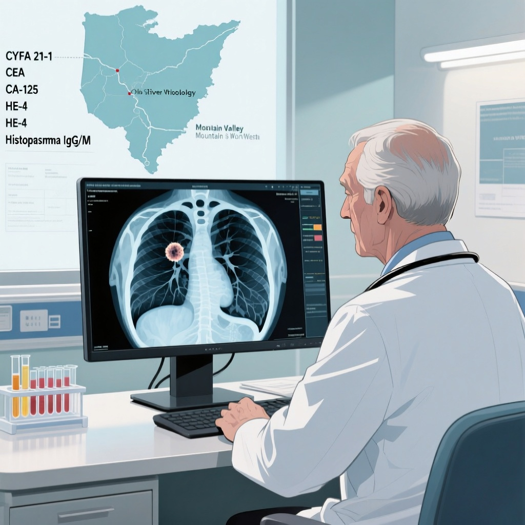
This investigation evaluated patients with 6-30 mm indeterminate pulmonary nodules from two geographically distinct US cohorts: the Ohio River Valley (ORV), where Histoplasma exposure is common, and the Mountain West (MW), a region with different background epidemiology. The inclusion of two cohorts is a notable design strength because it tests whether the model has value beyond a single endemic microenvironment.
The study included 366 patients overall: 282 from the ORV cohort and 84 from the MW cohort. For each patient, investigators calculated the Mayo score and measured serum concentrations of four commercial cancer protein biomarkers: CYFRA 21-1, carcinoembryonic antigen (CEA), CA-125, and human epididymis protein 4 (HE-4). They also measured Histoplasma IgG and IgM antibodies.
The primary analytic framework relied on area under the receiver operating characteristic curve, or AUC, with 95% confidence intervals. This metric reflects discrimination, meaning the ability of a model to correctly rank malignant nodules higher than benign nodules. The investigators assessed individual components, pairwise combinations, and a full multivariable logistic regression model that integrated clinical prediction, cancer biomarkers, and fungal serology. They also examined risk reclassification, an especially relevant outcome for pulmonary nodule clinics because management decisions often hinge on movement across low-, intermediate-, and high-risk thresholds.
The abstract does not provide a detailed breakdown of calibration, prespecified risk categories, prevalence of malignancy, histologic subtypes, or the final reference standard for all nodules. Those details will matter when the full article is interpreted, but the design as presented is appropriate for an initial diagnostic performance study.
Key Results
Performance of the Mayo model alone
The Mayo model showed moderate discrimination, with an AUC of 0.74 (95% CI, 0.67-0.80) in the ORV cohort and 0.68 (95% CI, 0.59-0.78) in the MW cohort. These results are clinically believable and consistent with longstanding experience: clinical-radiographic models are useful, but they leave meaningful uncertainty, particularly when the pretest landscape differs from the original derivation population.
Performance of biomarker combinations
When cancer biomarkers were combined with fungal antibodies, the AUC was 0.74 (95% CI, 0.68-0.80) in the ORV cohort and 0.76 (95% CI, 0.64-0.86) in the MW cohort. This is an interesting intermediate finding. Biomarkers alone or in limited combinations can approach the performance of a standard clinical model, suggesting that blood-based biologic data carry substantial diagnostic information.
However, the clinically most relevant result came from the full integrated model. When Mayo score, cancer protein markers, and Histoplasma antibodies were all included together, discrimination improved to an AUC of 0.80 (95% CI, 0.75-0.87) in the ORV cohort and 0.79 (95% CI, 0.70-0.88) in the MW cohort. In both cohorts, the combined strategy outperformed the Mayo model alone numerically. The gain appears modest in absolute AUC terms, but in pulmonary nodule management, even moderate improvements may meaningfully alter the downstream use of PET imaging, short-interval surveillance, transthoracic biopsy, bronchoscopy, or surgical resection.
Intermediate-risk nodules: the most clinically actionable subgroup
The most compelling part of the report is the effect on intermediate-risk nodules. These are the nodules that generate the greatest uncertainty and the widest variation in management. According to the abstract, the full model correctly upgraded malignancy risk in 46.3% of patients in one cohort and 42.9% in the other. Although the abstract specifically mentions upgrading rather than full bidirectional reclassification detail, these figures suggest that the integrated model can move a substantial fraction of otherwise ambiguous cases toward more decisive management.
That matters because intermediate-risk patients are exactly those for whom clinicians struggle with whether to observe, biopsy, or resect. A tool that better identifies malignancy in this group could reduce delays in diagnosis. Conversely, a combined model that recognizes fungal-serology-supported benign disease might also help avoid invasive procedures, particularly in endemic regions. The abstract emphasizes correct upgrading, but the full paper will be important to review for rates of correct downgrading and the balance of false positives versus false negatives.
Why the geography signal is important
The inclusion of both the ORV and MW cohorts deserves emphasis. In the ORV, Histoplasma exposure is a highly plausible benign mimic of cancer; in that setting, fungal antibodies may add specific contextual value. The persistence of benefit in the MW cohort suggests that the integrated approach may have broader utility, although the smaller sample size in MW produces wider confidence intervals and greater uncertainty. The fact that the full model still reached an AUC of 0.79 in the MW group supports the view that this is not merely a local epidemiologic artifact.
Clinical Interpretation
This study aligns with an increasingly important principle in pulmonary nodule evaluation: no single data stream is sufficient. Radiographic appearance, patient-level cancer risk, regional infectious epidemiology, and blood-based biomarkers all capture different facets of the same diagnostic problem. Integrating these sources can outperform any one component alone.
From a workflow perspective, the proposed model is attractive because it relies on commercially available cancer protein markers and relatively accessible fungal serologies. That gives the strategy near-term translational potential. A clinician evaluating a 12 mm spiculated upper-lobe nodule in a former smoker living in an endemic fungal region often faces conflicting signals. If a blood panel indicates low cancer biomarker burden but positive Histoplasma antibodies, the posterior probability of benign disease may shift meaningfully. Conversely, elevated cancer-associated proteins in a patient with equivocal imaging could justify expedited tissue diagnosis.
The study also highlights a broader limitation of conventional nodule prediction models: they are largely phenotype-based, not biology-based. They infer risk from age, smoking, and CT morphology, but they do not directly measure host or tumor biology. Biomarker integration is one route toward closing that gap. Although the markers used here are not lung cancer-specific in the strictest sense, a panel approach can still be informative if its added value is demonstrated in realistic populations.
Expert Commentary
There is strong biological plausibility behind the model. CYFRA 21-1 reflects cytokeratin 19 fragments and has been associated with non-small cell lung cancer, especially squamous histology. CEA remains a familiar though nonspecific tumor marker. CA-125 and HE-4 are more commonly discussed in gynecologic oncology but may reflect broader epithelial malignancy and inflammatory-tumor interactions. Histoplasma IgG and IgM antibodies, meanwhile, provide evidence of exposure or recent immune response to a fungal pathogen that can produce calcified or noncalcified nodules and granulomas. The conceptual innovation here is not that any one marker is definitive, but that the pattern across markers may refine probability estimates.
Current guideline frameworks, including those from the American College of Chest Physicians and other thoracic societies, generally recommend estimating pretest probability, then using imaging, surveillance, or tissue diagnosis according to risk thresholds. This study does not replace those pathways; rather, it proposes a more biologically informed way to assign the probability estimate that drives those decisions. If validated prospectively, such a model could be inserted upstream in the diagnostic algorithm, particularly for nodules that remain indeterminate after CT review.
Several limitations should temper interpretation. First, the abstract does not specify whether the model underwent external validation separate from model development within the two cohorts, nor does it provide calibration statistics. High discrimination alone is not enough; clinicians need to know whether predicted probabilities match observed outcomes. Second, the MW cohort is relatively small, which reduces precision. Third, some included biomarkers are nonspecific and may be influenced by comorbid conditions, inflammation, renal function, or other cancers. Fourth, Histoplasma serology performance depends on timing of exposure, immune status, and background seroprevalence. A positive antibody result does not prove that a nodule is benign or attributable to histoplasmosis.
Another important limitation is implementation detail. The abstract reports improved AUC and correct upgrading in intermediate-risk nodules, but it does not quantify decision-curve benefit, net reduction in procedures, or the impact on missed cancers. Those outcomes will determine clinical adoption more than AUC alone. A model can improve discrimination statistically yet fail to alter care in a net-beneficial way if threshold selection is poorly aligned with practice.
Implications for Practice
For pulmonologists, thoracic surgeons, oncologists, and radiologists, the immediate message is not that every nodule should undergo this exact panel tomorrow. Rather, the study suggests that multimodal blood testing may be most useful in patients whose estimated malignancy risk remains uncertain after standard assessment, particularly in areas where benign fungal disease is common.
Potential use cases include nodules in the intermediate pretest probability range, discordant cases with suspicious CT findings but low overall clinical risk, and patients in whom invasive testing carries higher procedural risk. In such scenarios, a biomarker-informed probability estimate could support shared decision-making. If future studies show that this approach safely reduces unnecessary biopsies or accelerates diagnosis of resectable cancers, it could become a practical adjunct in pulmonary nodule clinics and lung cancer screening programs.
However, clinicians should resist overinterpreting a positive or negative biomarker panel in isolation. These tests should be viewed as probability modifiers, not diagnostic verdicts. Integration with CT morphology, growth assessment, PET findings, smoking exposure, regional epidemiology, and patient preferences will remain essential.
Research Priorities
The next steps are clear. Prospective validation in larger, more diverse populations is needed, ideally with prespecified management thresholds and external calibration. Comparative studies against other modern nodule models would be valuable, especially those incorporating radiomics or machine learning. It will also be important to determine whether a reduced biomarker set could preserve most of the performance while simplifying cost and logistics.
Health economic analyses should follow. A modest improvement in AUC can still be highly valuable if it reduces unnecessary PET scans, bronchoscopies, transthoracic biopsies, or wedge resections. Conversely, widespread adoption would be difficult to justify if biomarker testing adds substantial cost without clearly improving patient-centered outcomes.
Finally, geographic tailoring deserves further study. Endemic fungal regions may benefit disproportionately from serology-enhanced models, but similar principles could be extended to other regional mimics of malignancy. The broader lesson is that pulmonary nodule diagnostics should account for local epidemiology rather than assuming a uniform national risk environment.
Conclusion
Holmes and colleagues provide evidence that integrating commercial cancer protein biomarkers and Histoplasma antibodies with the Mayo clinical prediction model improves discrimination of benign versus malignant indeterminate pulmonary nodules across two US regions. The strongest clinical signal appears in intermediate-risk nodules, where uncertainty is greatest and management consequences are most immediate.
This is not yet a definitive practice-changing study, but it is an important step toward biologically informed nodule assessment. In regions where fungal disease commonly complicates radiographic interpretation, adding serologic context to cancer biomarker data and standard prediction models is a rational and potentially useful strategy. The key question now is whether these statistical gains translate into fewer unnecessary invasive procedures, faster diagnosis of lung cancer, and better overall outcomes in prospective care pathways.
Funding and ClinicalTrials.gov
The abstract provided does not report funding information or a ClinicalTrials.gov registration number. Readers should consult the full Annals of Surgery publication for these details.
References
Holmes HM, McGann KC, Chen SC, Welch C, Zou Y, Antic S, Forero YJ, Argaw SA, Chen H, Kaizer A, Baron A, Meguid R, Maldonado F, Deppen SA, Grogan EL. Combining Commercial Cancer Protein Biomarkers and Benign Fungal Antibodies Improves Diagnostic Accuracy in Pulmonary Nodules. Ann Surg. 2026 Jun 10. PMID: 42265763.
Gould MK, Donington J, Lynch WR, Mazzone PJ, Midthun DE, Naidich DP, Wiener RS. Evaluation of individuals with pulmonary nodules: when is it lung cancer? Diagnosis and management of lung cancer, 3rd ed: American College of Chest Physicians evidence-based clinical practice guidelines. Chest. 2013;143(5 Suppl):e93S-e120S.
Swensen SJ, Silverstein MD, Ilstrup DM, Schleck CD, Edell ES. The probability of malignancy in solitary pulmonary nodules. Application to small radiologically indeterminate nodules. Arch Intern Med. 1997;157(8):849-855.
Mazzone PJ, Lam L. Evaluating the patient with a pulmonary nodule: a review. JAMA. 2022;327(3):264-273.
Wiener RS, Gould MK, Woloshin S, Schwartz LM, Clark JA. What do you mean, a spot? A qualitative analysis of patients’ reactions to discussions with their physicians about pulmonary nodules. Chest. 2013;143(3):672-677.