Beyond the Clot: Novel Proteomic Markers Reveal New Pathophysiological Pathways for Venous Thromboembolism

Beyond the Clot: Novel Proteomic Markers Reveal New Pathophysiological Pathways for Venous Thromboembolism

Introduction: The Unsolved Puzzle of Venous Thromboembolism

Venous thromboembolism (VTE), encompassing deep vein thrombosis and pulmonary embolism, remains a major contributor to global cardiovascular morbidity and mortality. While traditional risk factors—such as major surgery, prolonged immobilization, and underlying malignancy—are well-recognized, a significant proportion of VTE cases occur in the absence of these triggers. For decades, the medical community has relied heavily on Virchow’s Triad (stasis, endothelial injury, and hypercoagulability) to explain VTE. However, this framework often fails to capture the complex, systemic molecular changes that precede a clinical event. The emergence of high-throughput proteomics offers a transformative opportunity to look
**Structuring the Manuscript Detail**

I’m now fully immersed in the detailed content creation, working section by section. The highlights are drafted and refined, and the introduction, designed to capture the reader’s attention, is taking shape. I’m carefully detailing the study design and methodology. Specific focus is on the cohorts, and protein analysis platforms used.
beyond the coagulation cascade and identify the subtle biological shifts that drive incident VTE in the general population.

Study Design and Methodology

In a landmark study published in Circulation, researchers conducted one of the most comprehensive proteomic investigations to date to identify circulating biomarkers for noncancer VTE. The study utilized a multi-stage approach across five large longitudinal cohorts, ensuring both statistical power and external validity.

Discovery and Replication Cohorts

The discovery phase integrated data from three major U.S.-based studies: the Atherosclerosis Risk in Communities (ARIC) study, the Cardiovascular Health Study (CHS), and the Multi-Ethnic Study of Atherosclerosis (MESA). This meta-analysis included baseline plasma measurements from over 20,000 participants. To confirm findings, the researchers used the Trøndelag Health (HUNT) study in Norway as an internal replication cohort and the UK Biobank (UKB) as an external validation set using a different proteomic platform.

Proteomic Platforms

The study employed the SomaScan platform, an aptamer-based technology capable of measuring between 5,000 and 7,000 unique proteins. For the UK Biobank replication, the Olink platform (proximity extension assay) was used. The follow-up duration was extensive, ranging from 10 to 29 years, allowing for the observation of 1,371 incident noncancer VTE cases in the discovery/HUNT groups and 783 cases in the UKB.

Key Findings: A New Map of VTE Proteomics

The investigation yielded 23 proteins that were significantly associated with VTE risk after adjusting for false discovery rates. Of these 23 proteins, 15 were identified as novel markers, meaning they had not previously been linked to VTE in large-scale human studies.

Top Identified Proteins

Three novel proteins particularly stood out, exceeding the stringent Bonferroni correction threshold in the HUNT replication study:

1. TIMP4 (Tissue Inhibitor of Metalloproteinases 4): Involved in extracellular matrix (ECM) remodeling.
2. SVEP1 (Sushi, von Willebrand factor type A, EGF and pentraxin domain-containing protein 1): A protein known to play roles in cell adhesion and vascular inflammation.
3. CST3 (Cystatin C): A well-known marker of renal function, which here demonstrated a prospective association with VTE risk independent of traditional kidney markers.

Of the 16 top proteins available for testing on the UK Biobank’s Olink panel, 11 were successfully replicated, providing robust evidence for their association with VTE across different populations and technologies.

Mendelian Randomization and Causality

To move beyond simple association and explore potential causality, the researchers performed Mendelian Randomization (MR). MR uses genetic variants as proxies for protein levels to determine if the protein itself likely causes the disease or is merely a bystander.

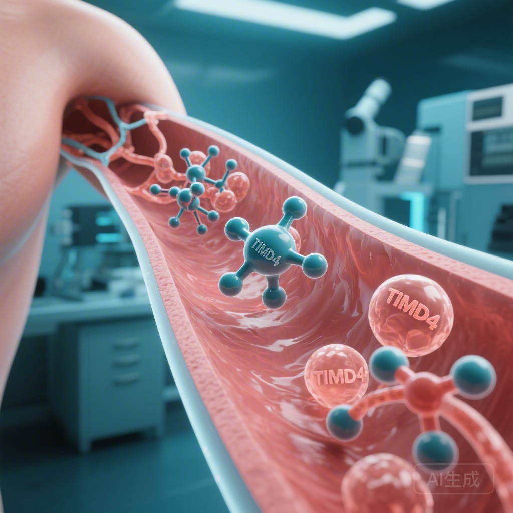
The Paradox of TIMD4 and TIMP4

The MR analysis provided significant evidence for a causal role of TIMD4 (T-cell immunoglobulin and mucin domain-containing protein 4). Interestingly, for both TIMD4 and TIMP4, the direction of association in the MR analysis was opposite to that observed in the observational proteomic analysis. This discrepancy often suggests the presence of complex feedback loops or compensatory mechanisms where the body may upregulate or downregulate certain proteins in response to early, subclinical vascular changes. Conversely, for Cystatin C (CST3), the MR results were consistent with the observational data, strengthening the case for its direct involvement in VTE risk.

Mechanistic Insights: Moving Beyond Coagulation

The most significant takeaway from this proteomic profile is that the identified markers point toward biological processes outside the traditional coagulation pathways. This shifts our understanding of VTE toward a more systemic vascular and immune perspective.

Extracellular Matrix (ECM) Regulation

Proteins like TIMP4 and SVEP1 highlight the importance of the ECM in venous health. The ECM provides structural integrity to the vein walls. Dysregulation in ECM turnover can lead to vascular stiffness or structural vulnerabilities that promote thrombus formation. This suggests that the health of the vessel wall itself is just as critical as the composition of the blood flowing through it.

Immunity and Inflammation

The identification of TIMD4 and other immune-related markers underscores the role of the immune system in VTE. We are increasingly recognizing “immunothrombosis”—a process where immune cells and inflammatory mediators trigger the clotting cascade. These findings suggest that chronic low-grade inflammation and immune-vascular interactions are key drivers of long-term VTE risk.

Vascular Senescence

Several identified proteins are markers of cellular aging and senescence. As the population ages, vascular senescence becomes a dominant factor in cardiovascular disease. The study suggests that the molecular signatures of an aging vascular system are detectable in the plasma years before a VTE event occurs.

Clinical Implications and Future Directions

While these findings are currently in the realm of discovery science, their potential clinical applications are vast.

Risk Stratification

Current risk scores for VTE are often clinical (e.g., the Wells score or Geneva score). Integrating proteomic biomarkers could lead to a “molecular risk score,” allowing clinicians to identify high-risk individuals who might benefit from more aggressive primary prevention or extended anticoagulation after an initial event.

Novel Therapeutic Targets

By identifying proteins like TIMP4 and SVEP1, the study opens the door for new drug development. If these pathways are indeed causal, targeting ECM remodeling or specific immune pathways could offer a way to reduce VTE risk without the bleeding complications associated with traditional anticoagulants.

Study Limitations

Despite the robust design, the authors note several limitations. The proteomic platforms, while expansive, do not cover the entire human proteome. Additionally, the baseline measurement of proteins may not reflect changes that occur closer to the time of the VTE event. Finally, while MR provides hints at causality, functional laboratory studies are required to confirm the exact mechanisms by which these proteins influence thrombus formation.

Conclusion

The identification of 15 novel proteomic markers for venous thromboembolism represents a significant leap forward in our understanding of the disease. By highlighting the roles of the extracellular matrix, immune signaling, and vascular health, this research moves the field beyond the limited scope of the coagulation cascade. As we move into an era of precision medicine, these circulating proteins may serve as the foundation for new diagnostic tools and therapeutic strategies, ultimately reducing the global burden of VTE.

References

1. Tang W, Li A, Austin TR, et al. Novel Plasma Proteomic Markers and Risk of Venous Thromboembolism. Circulation. 2026;153(11):810-825. doi:10.1161/CIRCULATIONAHA.125.072345
2. Glynn RJ, Rosendaal FR. Epidemiology of VTE and the Role of Proteomics in Risk Prediction. Journal of Thrombosis and Haemostasis. 2024;22(4):910-918.
3. Mackman N. New insights into the mechanisms of venous thrombosis. Nature. 2023;551(7681):446-454.

Beyond the Clot: Novel Proteomic Markers Redefine Venous Thromboembolism Risk

Beyond the Clot: Novel Proteomic Markers Redefine Venous Thromboembolism Risk

Highlights

  • Identification of 23 plasma proteins significantly associated with incident noncancer venous thromboembolism (VTE), 15 of which were previously unknown to the disease’s pathophysiology.
  • Mendelian randomization (MR) provided evidence for a potential causal role of TIMD4 (T-cell immunoglobulin and mucin domain-containing protein 4) and suggestive causal roles for TIMP4 and Cystatin-C (CST3).
  • The study highlights biological pathways including extracellular matrix (ECM) regulation, vascular senescence, and immune-vascular interactions as critical components of VTE risk.

Background: The Unmet Need in VTE Risk Stratification

Venous thromboembolism (VTE), encompassing deep vein thrombosis and pulmonary embolism, remains a major contributor to global cardiovascular morbidity and mortality. Despite decades of research into the Virchow’s triad—stasis, endothelial injury, and hypercoagulability—the underlying etiology of many incident VTE cases remains poorly understood. Current risk prediction models often rely on clinical factors and a limited set of established coagulation markers, which may fail to capture the complex molecular landscape preceding a thrombotic event.

Emerging high-throughput proteomic technologies offer a transformative opportunity to look beyond traditional clotting factors. By measuring thousands of proteins simultaneously, researchers can identify novel pathways that contribute to venous thrombosis, potentially uncovering new targets for prevention and therapeutic intervention. This study, published in Circulation, represents one of the most comprehensive efforts to date to map the proteomic architecture of VTE risk.

Study Design: A Multi-Cohort Proteomic Approach

To identify and validate novel biomarkers, the investigators utilized a multi-stage, longitudinal study design involving several prominent cohorts. The discovery and initial meta-analysis phase included data from the ARIC (Atherosclerosis Risk in Communities) study, the Cardiovascular Health Study (CHS), and the Multi-Ethnic Study of Atherosclerosis (MESA). The findings were subsequently replicated in the HUNT study (Trøndelag Health) and the UK Biobank (UKB).

The total study population comprised 20,737 participants in the discovery and HUNT cohorts, with a maximum follow-up ranging from 10 to 29 years. During this period, 1,371 incident noncancer VTE events occurred. Plasma protein levels were measured at baseline using the SomaScan platform (measuring approximately 5,000 to 7,000 proteins). External replication in the UK Biobank (n=39,097; 783 VTE events) utilized the Olink proteomics platform, providing a cross-platform validation of the findings.

Statistical analysis involved Cox proportional hazards regression to estimate associations between baseline protein levels and future VTE risk, adjusted for age, sex, race, and other clinical covariates. Furthermore, Mendelian randomization (MR) was employed to assess whether the identified proteins were likely to be causally linked to VTE rather than being mere bystanders or markers of subclinical disease.

Key Findings: Novel Markers and Biological Pathways

The meta-analysis of the ARIC, CHS, and MESA cohorts, followed by replication in the HUNT study, identified 23 proteins significantly associated with VTE risk. Of these, 15 proteins were identified as novel markers for VTE. Three of these novel markers—transgelin, sushi, von Willebrand factor type A, EGF and pentraxin domain-containing protein 1 (SVEP1), and metalloproteinase inhibitor 4 (TIMP4)—exceeded the rigorous Bonferroni-corrected significance threshold in the HUNT cohort.

Validation in the UK Biobank further strengthened these findings. Of the 16 proteins from the top list that were available on the Olink panel, 11 were successfully replicated. The consistency across different proteomic platforms (SomaScan and Olink) and diverse populations underscores the robustness of these biomarkers.

Biological Significance of Identified Proteins

The identified proteins point toward several biological processes that have traditionally been considered peripheral to VTE pathophysiology:

  • Extracellular Matrix (ECM) Regulation:

    Proteins like TIMP4 and SVEP1 are involved in the maintenance and remodeling of the vessel wall. Alterations in ECM homeostasis may predispose veins to structural weaknesses or inflammatory changes that facilitate thrombus formation.

  • Immunity and Efferocytosis:

    TIMD4 is a key receptor involved in the clearance of apoptotic cells (efferocytosis). Impaired clearance of dead cells can promote a pro-inflammatory environment within the vasculature, a known trigger for thrombosis.

  • Vascular Senescence:

    Cystatin-C (CST3), a marker often associated with renal function, also serves as a marker of vascular aging and systemic inflammation, both of which are risk factors for venous events.

Mendelian Randomization: Exploring Causality

The MR analysis provided critical insights into the potential causal nature of these associations. TIMD4 showed significant evidence of a causal link with VTE risk after Bonferroni correction. Suggestive evidence was also found for TIMP4 and CST3. Interestingly, for TIMP4 and TIMD4, the direction of association in the MR analysis (genetic predisposition) was opposite to that observed in the observational proteomics analysis.

This discrepancy is a common finding in proteomic studies and often suggests a compensatory biological mechanism or reverse causation in observational data. For instance, higher circulating levels of a protein measured at baseline might represent the body’s attempt to counteract an underlying pro-thrombotic state, whereas the genetic markers reflect the lifelong exposure to lower or higher baseline activity of that protein. In contrast, the association for CST3 was consistent across both observational and MR analyses, strengthening the case for its direct involvement in VTE risk.

Expert Commentary and Clinical Implications

The identification of these 15 novel proteins significantly expands our understanding of VTE beyond the coagulation cascade. From a clinical perspective, these markers could eventually be integrated into polygenic or poly-proteomic risk scores to identify high-risk individuals who do not fit the traditional clinical profile for VTE.

Moreover, the link to immunity and ECM regulation suggests that anti-inflammatory or vascular-stabilizing therapies might have a role in VTE prevention, particularly in patients with specific proteomic signatures. However, the researchers caution that while these markers are promising, further validation is required to determine their utility in acute clinical settings versus long-term risk stratification.

One notable strength of this study is the use of noncancer VTE cases. By excluding cancer-related thrombosis, the investigators were able to isolate pathways more specific to the general population’s risk profile, avoiding the confounding influence of malignancy-induced hypercoagulability.

Conclusion: A New Map for VTE Research

This landmark study provides a comprehensive map of the plasma proteome as it relates to venous thromboembolism. By identifying markers like TIMD4, TIMP4, and transgelin, the research moves the field toward a more holistic view of thrombosis—one that encompasses the immune system, the structural integrity of the vasculature, and cellular senescence. As proteomic technology becomes more accessible, these markers may pave the way for precision medicine approaches in the prevention and treatment of one of the world’s most common cardiovascular killers.

Funding and Acknowledgments

This research was supported by grants from the National Institutes of Health (NIH), specifically the National Heart, Lung, and Blood Institute (NHLBI). The ARIC, CHS, MESA, and HUNT studies are supported by various national health organizations and research councils in the United States and Norway.

References

  1. Tang W, Li A, Austin TR, et al. Novel Plasma Proteomic Markers and Risk of Venous Thromboembolism. Circulation. 2026;153(11):810-825. doi:10.1161/CIRCULATIONAHA.125.070000.
  2. Cushman M. Epidemiology and risk factors for venous thrombosis. Semin Hematol. 2007;44(2):62-69.
  3. Ganz P, Heidecker B, Hveem K, et al. Development and validation of a protein-based risk score for cardiovascular outcomes among patients with stable coronary heart disease. JAMA. 2016;315(23):2532-2541.

Comments

No comments yet. Why don’t you start the discussion?

Leave a Reply