Highlights
- Curcumin significantly reduces oxidative stress markers and apoptosis in methotrexate-induced ovarian injury.
- Antioxidant enzyme levels (SOD, CAT) increase, while MDA (a marker of lipid peroxidation) decreases with curcumin.
- Follicular reserve is preserved, with increased primordial follicle count and reduced atretic follicles.
- P53 and PARP-1 expression are modulated, suggesting a mechanistic link between curcumin’s antioxidant effects and apoptosis regulation.
Study Background and Disease Burden
Methotrexate (MTX) is a cornerstone in the management of various neoplastic (e.g., leukemia, lymphoma) and non-neoplastic diseases (e.g., rheumatoid arthritis, ectopic pregnancy). Despite its clinical efficacy, MTX is notorious for its off-target toxicity, particularly oxidative damage to rapidly dividing tissues such as the ovaries. Ovarian toxicity manifests as diminished follicle reserve, increased follicular atresia, and impaired reproductive lifespan — a significant concern for young women undergoing chemotherapy or chronic MTX therapy. There is a pressing need for effective strategies to mitigate chemotherapy-induced gonadotoxicity without compromising antitumor efficacy.
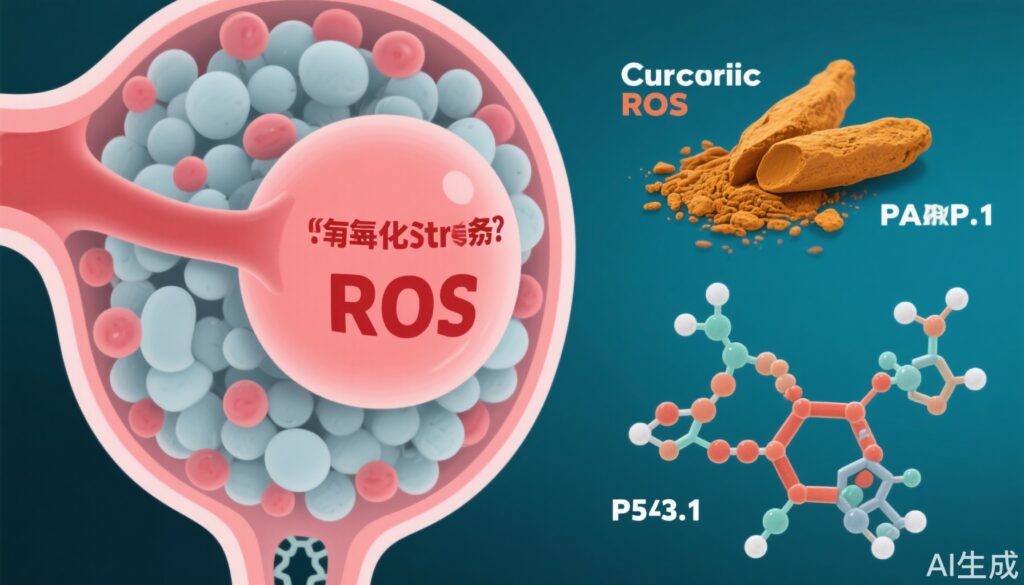
Curcumin, a polyphenolic compound derived from turmeric (Curcuma longa), is recognized for its potent antioxidant, anti-inflammatory, and antiapoptotic properties. Preclinical studies suggest curcumin’s potential to counteract various forms of tissue injury, but its role in preventing MTX-induced ovarian damage, especially via modulation of apoptosis-related pathways such as PARP-1 and P53, remains to be fully elucidated.
Study Design
This controlled animal study involved thirty-two young adult female Wistar albino rats, randomized into four groups (n=8 per group):
1. Control: vehicle only
2. Curcumin (Cur): 200 mg/kg/day
3. Methotrexate (MTX): 0.35 mg/kg/day
4. MTX + Curcumin: both agents for 28 days
After 28 days, ovarian tissue and serum were collected. The primary endpoints included biochemical markers of oxidative stress (SOD, CAT, MDA), anti-Müllerian hormone (AMH) as a marker of ovarian reserve, histological follicle counts (primordial, secondary, atretic follicles), and immunohistochemical analysis of P53 and PARP-1 expression.
Key Findings
Oxidative Stress and Antioxidant Enzymes:
– MTX administration resulted in a significant increase in MDA levels (a marker of lipid peroxidation and oxidative damage) compared to controls (p < 0.05).
– Curcumin co-treatment (MTX+Cur group) significantly reduced MDA levels relative to MTX alone (p < 0.05), indicating mitigation of oxidative stress.
– Antioxidant enzyme activities (SOD, CAT) were significantly higher in the MTX+Cur group than in the MTX group (p < 0.05 for both), reinforcing curcumin’s antioxidant action.
Ovarian Follicle Reserve and Morphology:
– The MTX-only group displayed a marked increase in atretic (degenerating) follicle count and a reduction in primordial follicle count, consistent with follicle loss and ovarian insufficiency.
– In contrast, the MTX+Cur group showed a significant decrease in atretic follicles (p < 0.05) and a notable increase in primordial follicles (p < 0.01) compared to MTX alone, suggesting preservation of follicle pool.
– Secondary follicle count and AMH levels were higher in MTX-treated groups, reflecting possible compensatory folliculogenesis, but these parameters were better preserved with curcumin co-administration.
Apoptosis Markers (P53 and PARP-1):
– Immunohistochemistry revealed a significant upregulation of P53 and PARP-1 in the MTX group (p < 0.05), indicating increased DNA damage and apoptosis.
– Curcumin treatment prevented this upregulation, supporting the hypothesis that curcumin modulates apoptotic pathways in addition to oxidative stress.
Statistical and Biological Significance:
– All key results reached statistical significance (p-values ranging from <0.05 to <0.01).
– The data provide compelling evidence that curcumin’s dual antioxidant and antiapoptotic actions can attenuate MTX-induced ovarian injury.
Expert Commentary
The findings of Keçeci and Karaoluk add to the growing body of preclinical evidence supporting curcumin as a candidate for fertility preservation in the context of chemotherapeutic regimens. The study’s robust design, including multiple biochemical, histological, and molecular endpoints, strengthens the translational relevance of the results. Notably, the modulation of P53 and PARP-1 — both central mediators of apoptosis and DNA repair — provides a mechanistic framework for curcumin’s protective effect that could be targeted in future drug development.
However, several limitations should be acknowledged. The rat model, while informative, may not fully capture the complexity of human ovarian physiology or drug metabolism. The high doses of curcumin used in animal studies are often not directly translatable to humans due to poor oral bioavailability. Additionally, the potential interaction of curcumin with the antitumor effects of MTX warrants further investigation, particularly in cancer-bearing models or clinical trials.
Clinical guidelines for fertility preservation in oncology currently prioritize established methods such as oocyte or embryo cryopreservation. Nonetheless, adjunctive pharmacological agents that are safe, accessible, and non-intrusive could significantly broaden the options for young women at risk of iatrogenic ovarian failure.
Conclusion
Curcumin demonstrates significant promise in reducing MTX-induced ovarian damage through its antioxidant and antiapoptotic effects, specifically via downregulation of P53 and PARP-1 pathways. These findings justify further translational studies to evaluate curcumin as an adjuvant for fertility preservation during chemotherapy, with careful attention to dosage, bioavailability, and safety in humans. Until human data are available, these results should be viewed as hypothesis-generating but highly encouraging for future clinical research.
References
1. Keçeci M, Karaoluk N. Effect of curcumin on methotrexate-induced ovarian damage and follicle reserve in rats: the role of PARP-1 and P53. Ann Med. 2025 Dec;57(1):2446688. doi: 10.1080/07853890.2024.2446688 IF: 4.3 Q1 . Epub 2024 Dec 27. PMID: 39729361 IF: 4.3 Q1 ; PMCID: PMC11702994 IF: 4.3 Q1 .
2. Practice Committee of the American Society for Reproductive Medicine. Fertility preservation in patients undergoing gonadotoxic therapy or gonadectomy: a committee opinion. Fertil Steril. 2019;112(6):1022-1033.
3. Hewlings SJ, Kalman DS. Curcumin: A Review of Its’ Effects on Human Health. Foods. 2017;6(10):92. doi:10.3390/foods6100092 IF: 5.1 Q1 .
4. Lambert JD, Elias RJ. The antioxidant and pro-oxidant activities of green tea polyphenols: a role in cancer prevention. Arch Biochem Biophys. 2010;501(1):65-72.